Cambridge A Level Biology 9700 — 2018 Feb/March Paper 4 · Variant 2
9700/42/F/M/18 · 10 questions · 100 marks · ≈113 min
The question paper and its mark scheme, free to read here and free to download. This is Cambridge’s own paper, exactly as it was sat.
Question paper28 pages
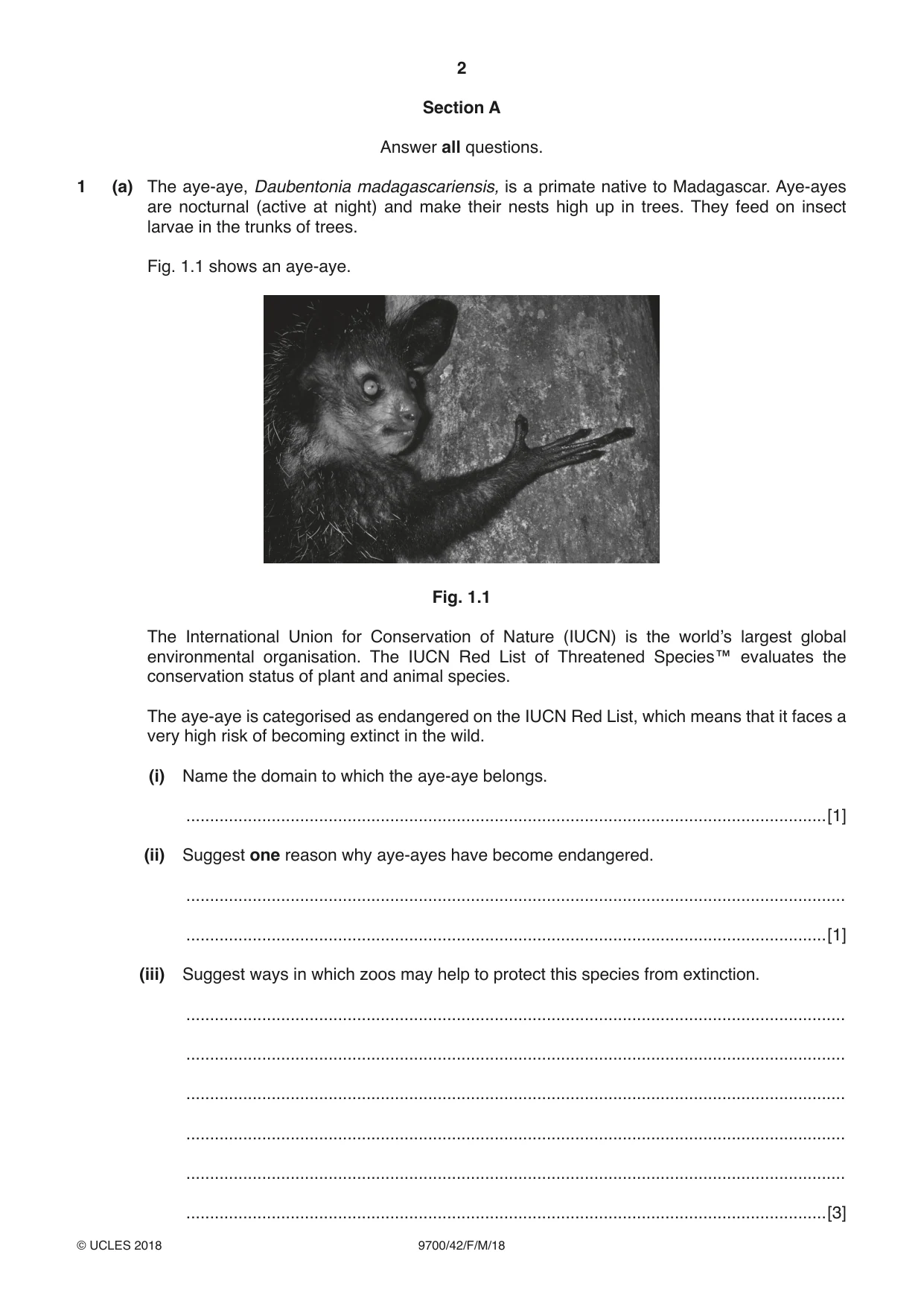

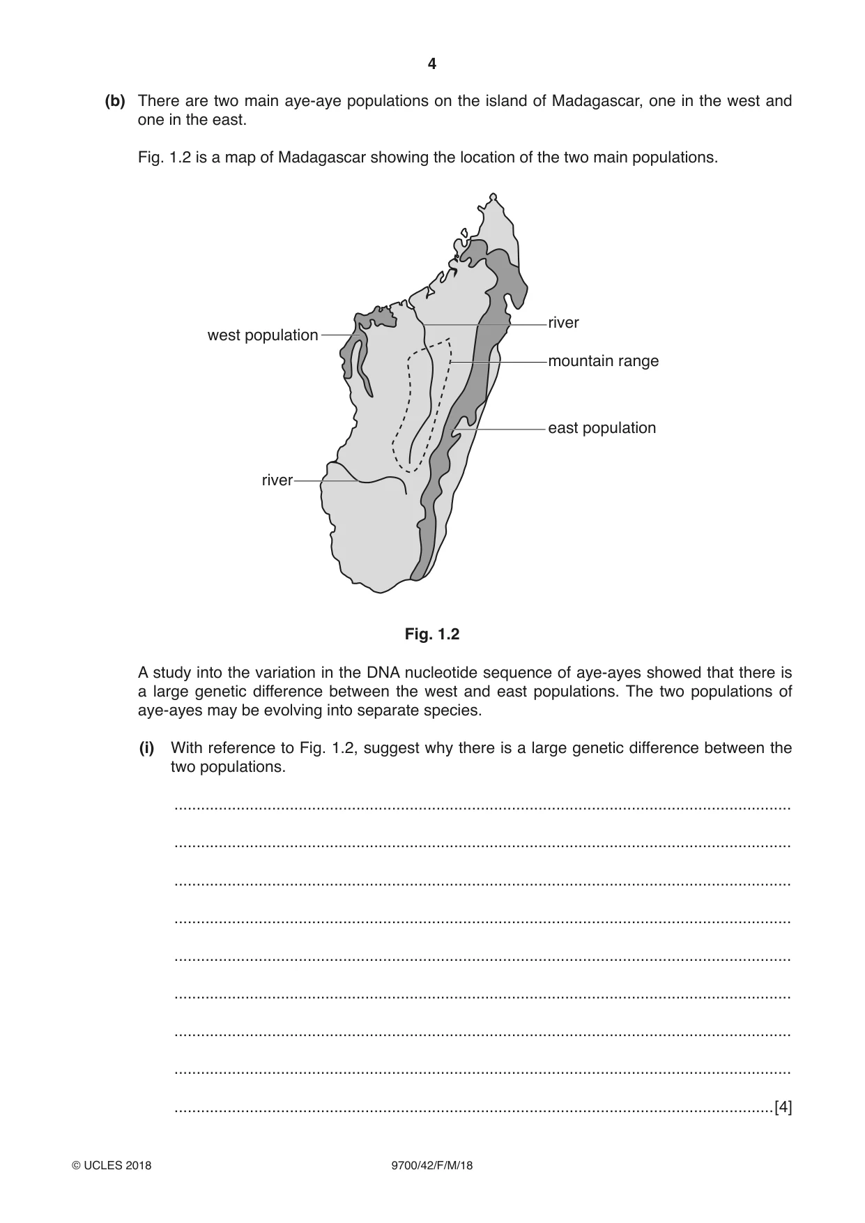




Mark scheme22 pages
Answers below. Sit the paper first if you are practising.
Questions as text
Q1 · The aye-aye, Daubentonia madagascariensis, is a primate native to Madagascar
1 (a) The aye-aye, Daubentonia madagascariensis, is a primate native to Madagascar. Aye-ayes are nocturnal (active at night) and make their nests high up in trees. They feed on insect larvae in the trunks of trees. Fig. 1.1 shows an aye-aye. Fig. 1.1 The International Union for Conservation of Nature (IUCN) is the world’s largest global environmental organisation. The IUCN Red List of Threatened Species™ evaluates the conservation status of plant and animal species. The aye-aye is categorised as endangered on the IUCN Red List, which means that it faces a very high risk of becoming extinct in the wild. (i) Name the domain to which the aye-aye belongs. .......................................................................................................................................[1] (ii) Suggest one reason why aye-ayes have become endangered. ........................................................................................................................................... .......................................................................................................................................[1] (iii) Suggest ways in which zoos may help to protect this species from extinction. ........................................................................................................................................... ........................................................................................................................................... ........................................................................................................................................... ........................................................................................................................................... ........................................................................................................................................... .......................................................................................................................................[3] Question 1 continues on page 4 (b) There are two main aye-aye populations on the island of Madagascar, one in the west and one in the east. Fig. 1.2 is a map of Madagascar showing the location of the two main populations. river west population mountain range east population river Fig. 1.2 A study into the variation in the DNA nucleotide sequence of aye-ayes showed that there is a large genetic difference between the west and east populations. The two populations of aye-ayes may be evolving into separate species. (i) With reference to Fig. 1.2, suggest why there is a large genetic difference between the two populations. ........................................................................................................................................... ........................................................................................................................................... ........................................................................................................................................... ........................................................................................................................................... ........................................................................................................................................... ........................................................................................................................................... ........................................................................................................................................... ........................................................................................................................................... .......................................................................................................................................[4] (ii) Name the type of speciation that may be occurring. ........................................................................................................................................... .......................................................................................................................................[1] (iii) Suggest and explain a pre-zygotic isolating mechanism that could prevent successful reproduction between aye-ayes of the two populations. ........................................................................................................................................... ........................................................................................................................................... ........................................................................................................................................... ........................................................................................................................................... ........................................................................................................................................... ........................................................................................................................................... ........................................................................................................................................... .......................................................................................................................................[2] [Total: 12]
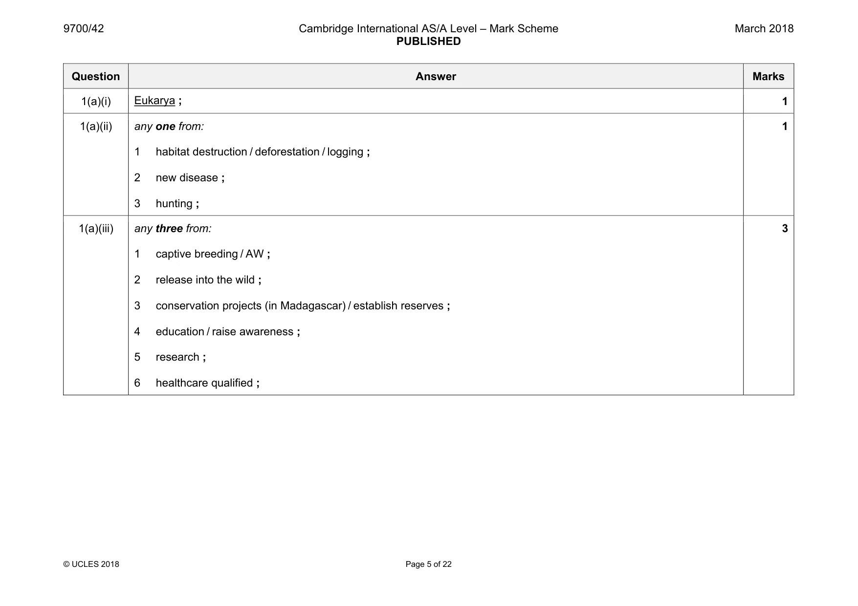
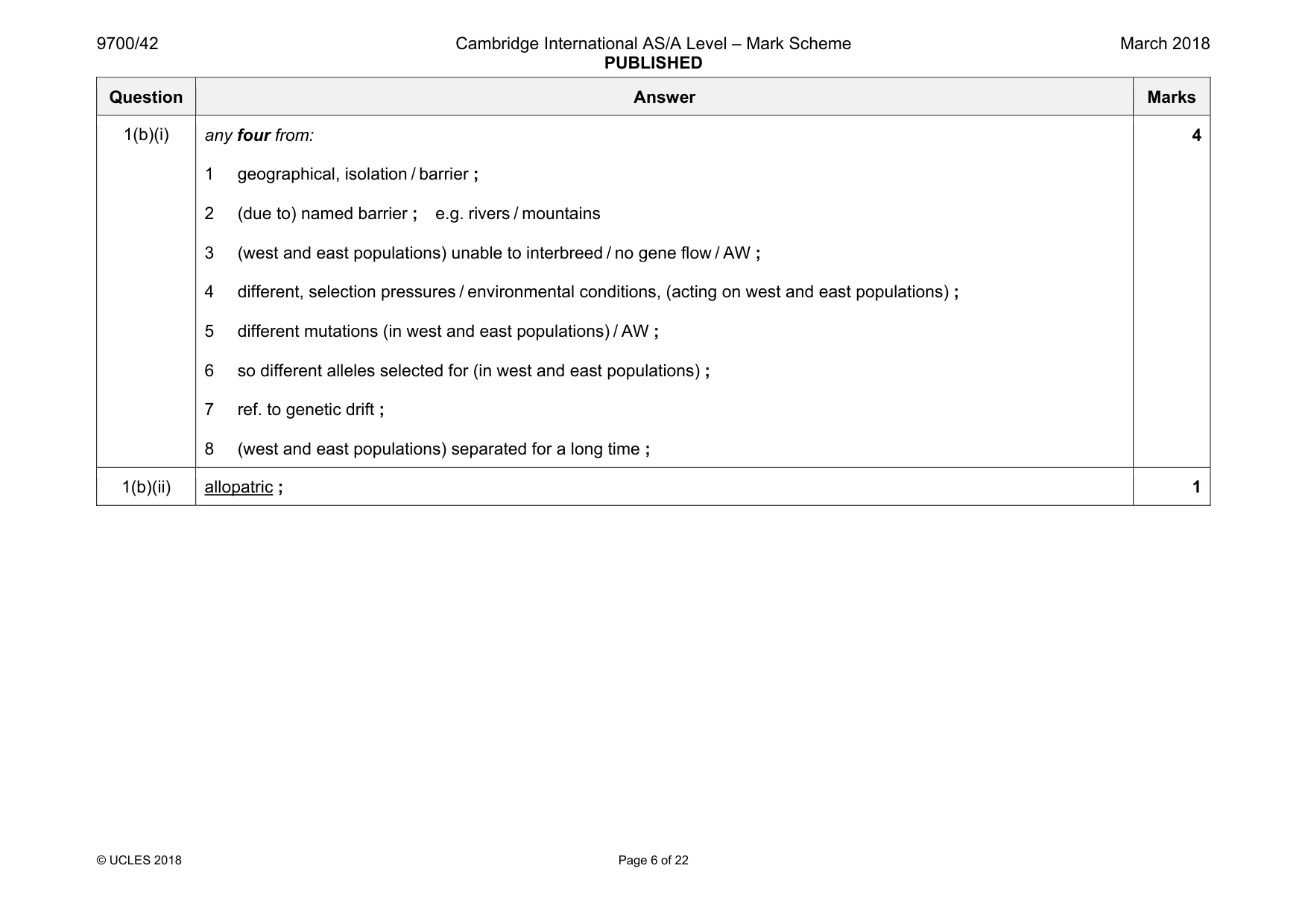
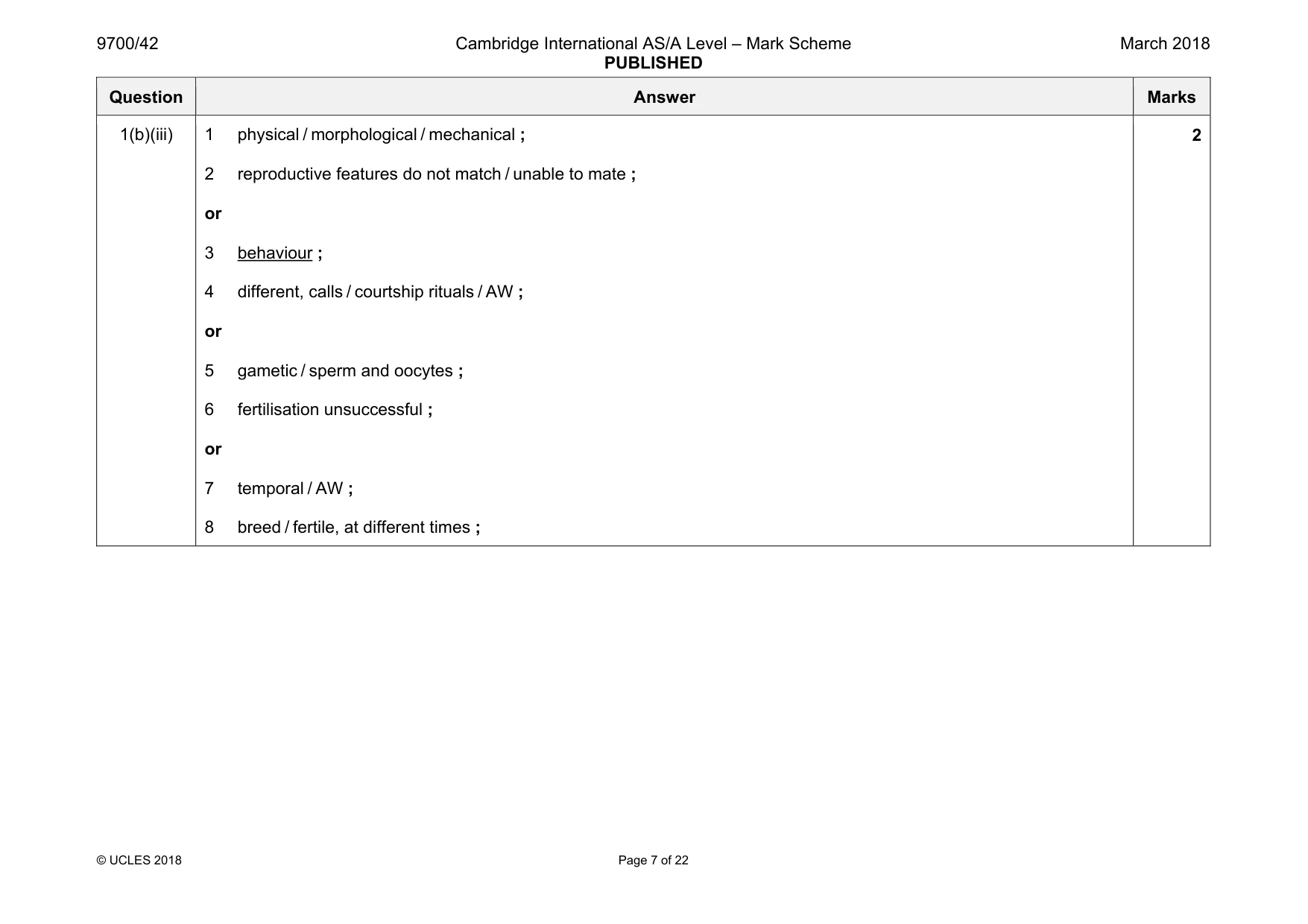
Mark scheme: 1(a)(i) 1 1(a)(ii) any one from: 1 habitat destruction / deforestation / logging ; 2 new disease ; 3 hunting ; 1 1(a)(iii) any three from: 1 captive breeding / AW ; 2 release into the wild ; 3 conservation projects (in Madagascar) / establish reserves ; 4 education / raise awareness ; 5 research ; 6 healthcare qualified ; 3 Question Answer Marks 1(b)(i) any four from: 1 geographical, isolation / barrier ; 2 (due to) named barrier ; e.g. rivers / mountains 3 (west and east populations) unable to interbreed / no gene flow / AW ; 4 different, selection pressures / environmental conditions, (acting on west and east populations) ; 5 different mutations (in west and east populations) / AW ; 6 so different alleles selected for (in west and east populations) ; 7 ref. to genetic drift ; 8 (west and east populations) separated for a long time ; 4 1(b)(ii) allopatric ; 1 Question Answer Marks 1(b)(iii) 1 physical / morphological / mechanical ; 2 reproductive features do not match / unable to mate ; or 3 behaviour ; 4 different, calls / courtship rituals / AW ; or 5 gametic / sperm and oocytes ; 6 fertilisation unsuccessful ; or 7 temporal / AW ; 8 breed / fertile, at different times ; 2
Q2 · Motor neurones are cells within the nervous system
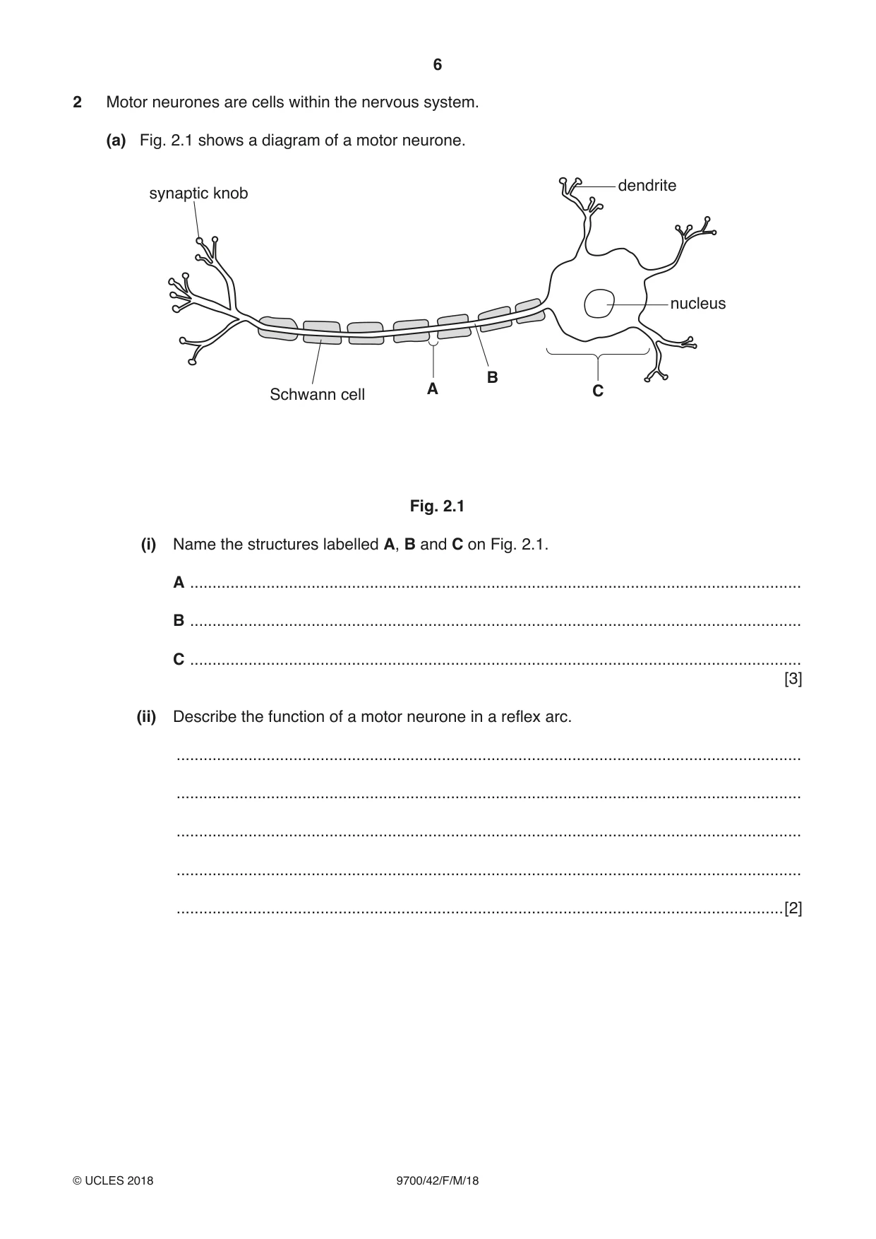
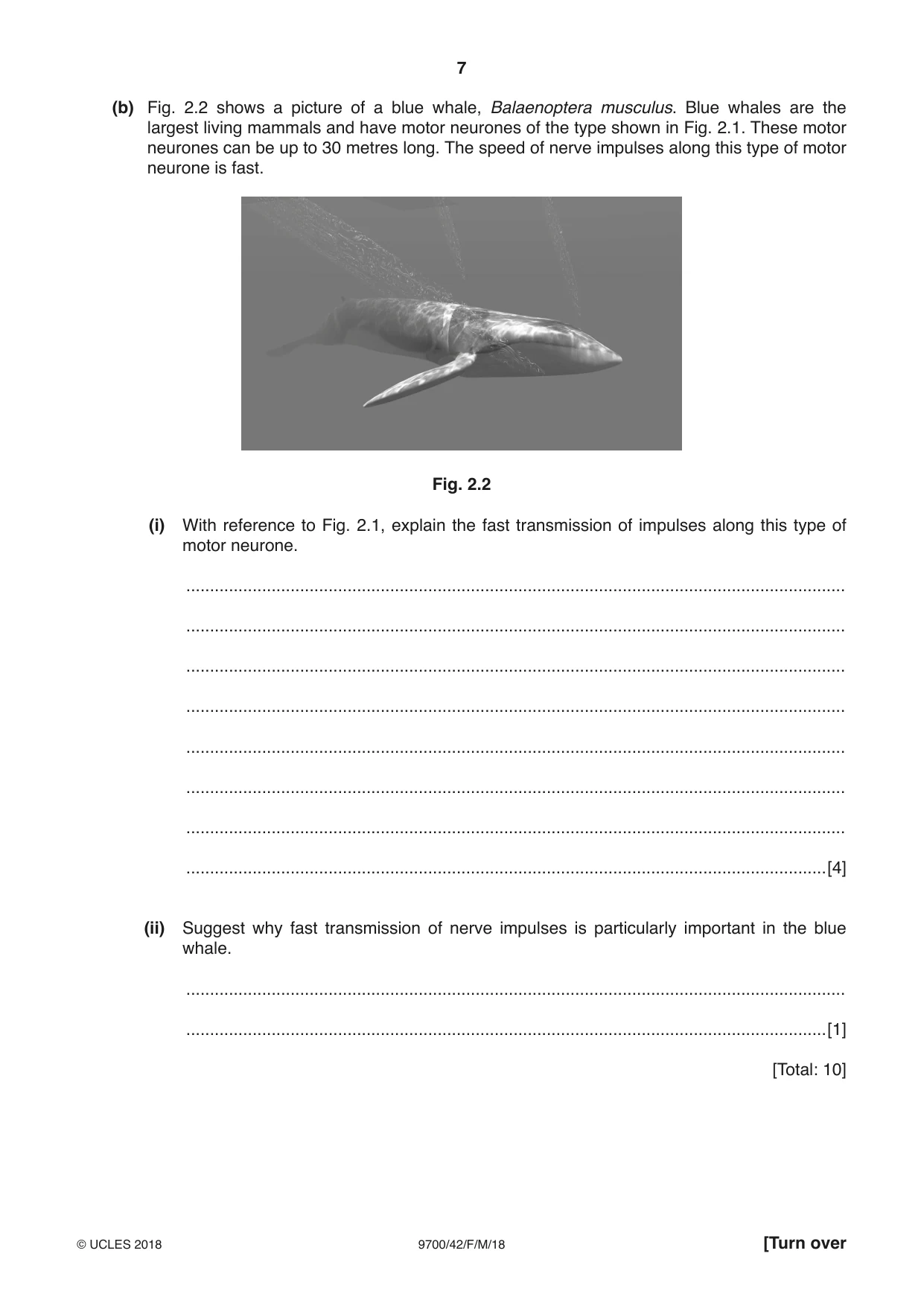
2 Motor neurones are cells within the nervous system. (a) Fig. 2.1 shows a diagram of a motor neurone. dendrite synaptic knob nucleus B A C Schwann cell Fig. 2.1 (i) Name the structures labelled A, B and C on Fig. 2.1. A ........................................................................................................................................ B ........................................................................................................................................ C ........................................................................................................................................ [3] (ii) Describe the function of a motor neurone in a reflex arc. ........................................................................................................................................... ........................................................................................................................................... ........................................................................................................................................... ........................................................................................................................................... .......................................................................................................................................[2] (b) Fig. 2.2 shows a picture of a blue whale, Balaenoptera musculus. Blue whales are the largest living mammals and have motor neurones of the type shown in Fig. 2.1. These motor neurones can be up to 30 metres long. The speed of nerve impulses along this type of motor neurone is fast. Fig. 2.2 (i) With reference to Fig. 2.1, explain the fast transmission of impulses along this type of motor neurone. ........................................................................................................................................... ........................................................................................................................................... ........................................................................................................................................... ........................................................................................................................................... ........................................................................................................................................... ........................................................................................................................................... ........................................................................................................................................... .......................................................................................................................................[4] (ii) Suggest why fast transmission of nerve impulses is particularly important in the blue whale. ........................................................................................................................................... .......................................................................................................................................[1] [Total: 10]
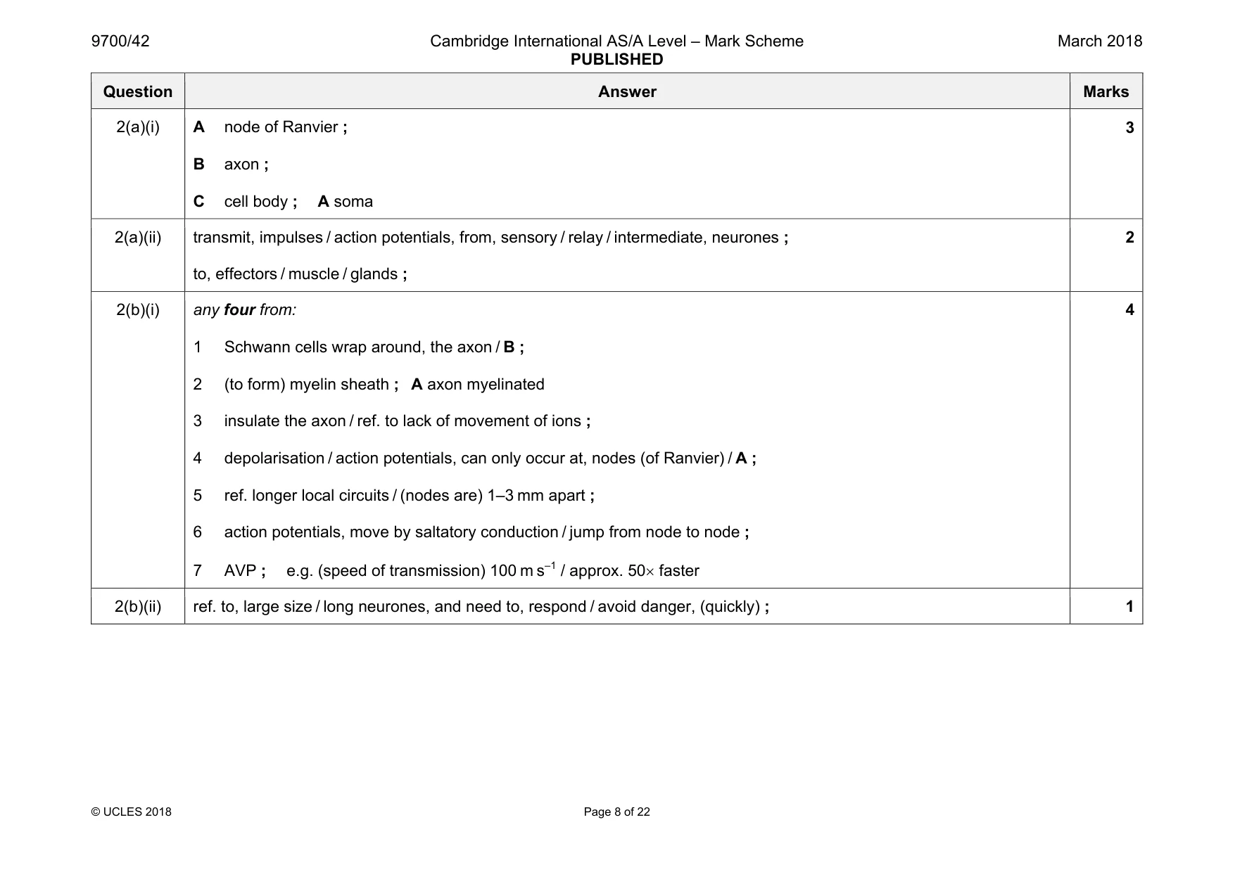
Mark scheme: 2(a)(i) A node of Ranvier ; B axon ; C cell body ; A soma 3 2(a)(ii) transmit, impulses / action potentials, from, sensory / relay / intermediate, neurones ; to, effectors / muscle / glands ; 2 2(b)(i) any four from: 1 Schwann cells wrap around, the axon / B ; 2 (to form) myelin sheath ; A axon myelinated 3 insulate the axon / ref. to lack of movement of ions ; 4 depolarisation / action potentials, can only occur at, nodes (of Ranvier) / A ; 5 ref. longer local circuits / (nodes are) 1–3 mm apart ; 6 action potentials, move by saltatory conduction / jump from node to node ; 7 AVP ; e.g. (speed of transmission) 100 m s–1 / approx. 50× faster 4 2(b)(ii) ref. to, large size / long neurones, and need to, respond / avoid danger, (quickly) ; 1
Q3 · The β-globin gene codes for the β-globin polypeptide of haemoglobin
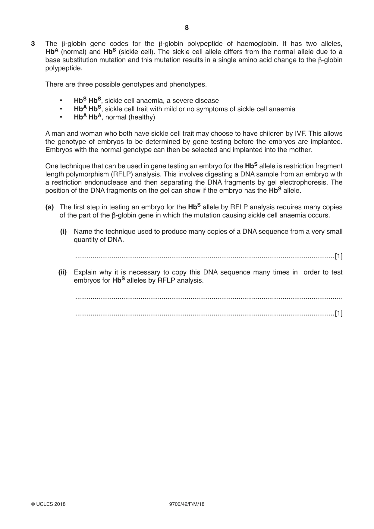
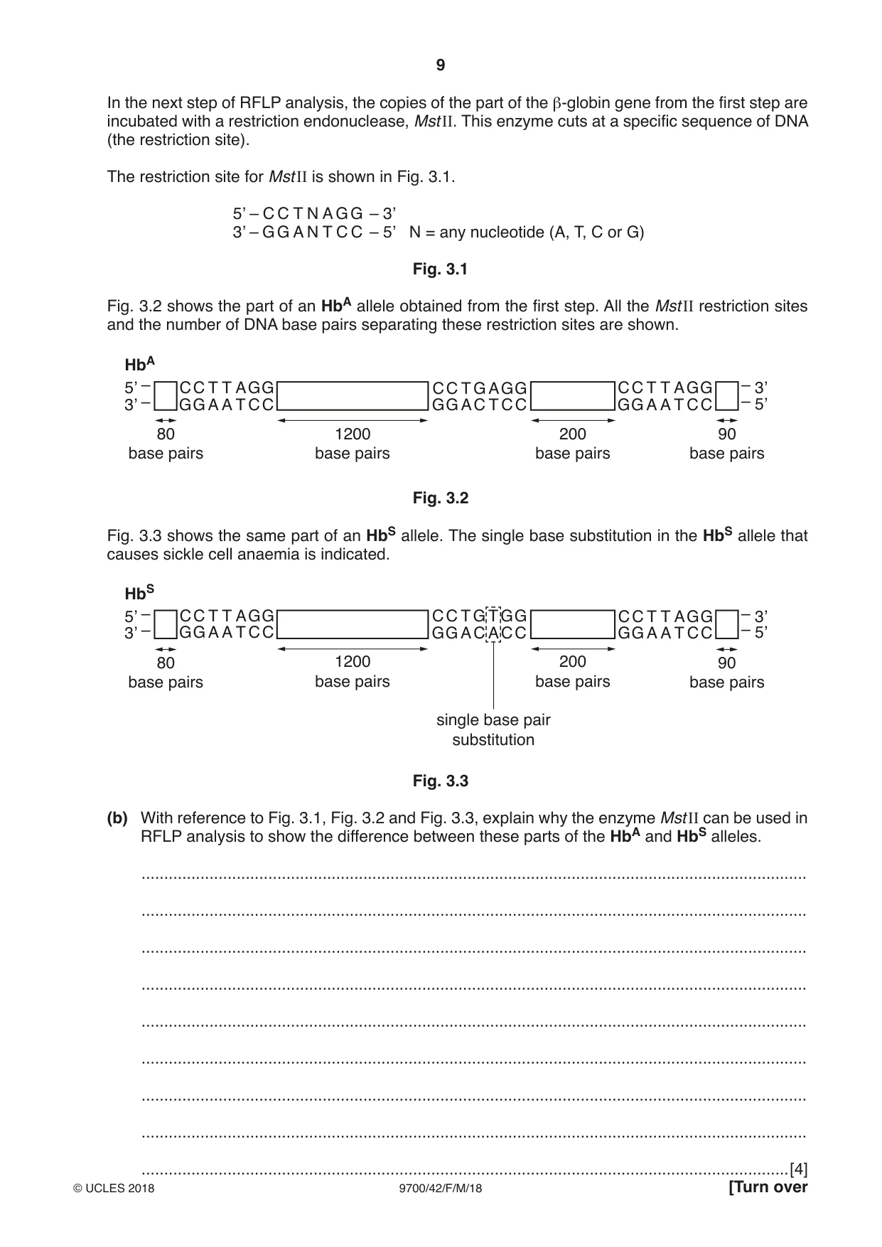
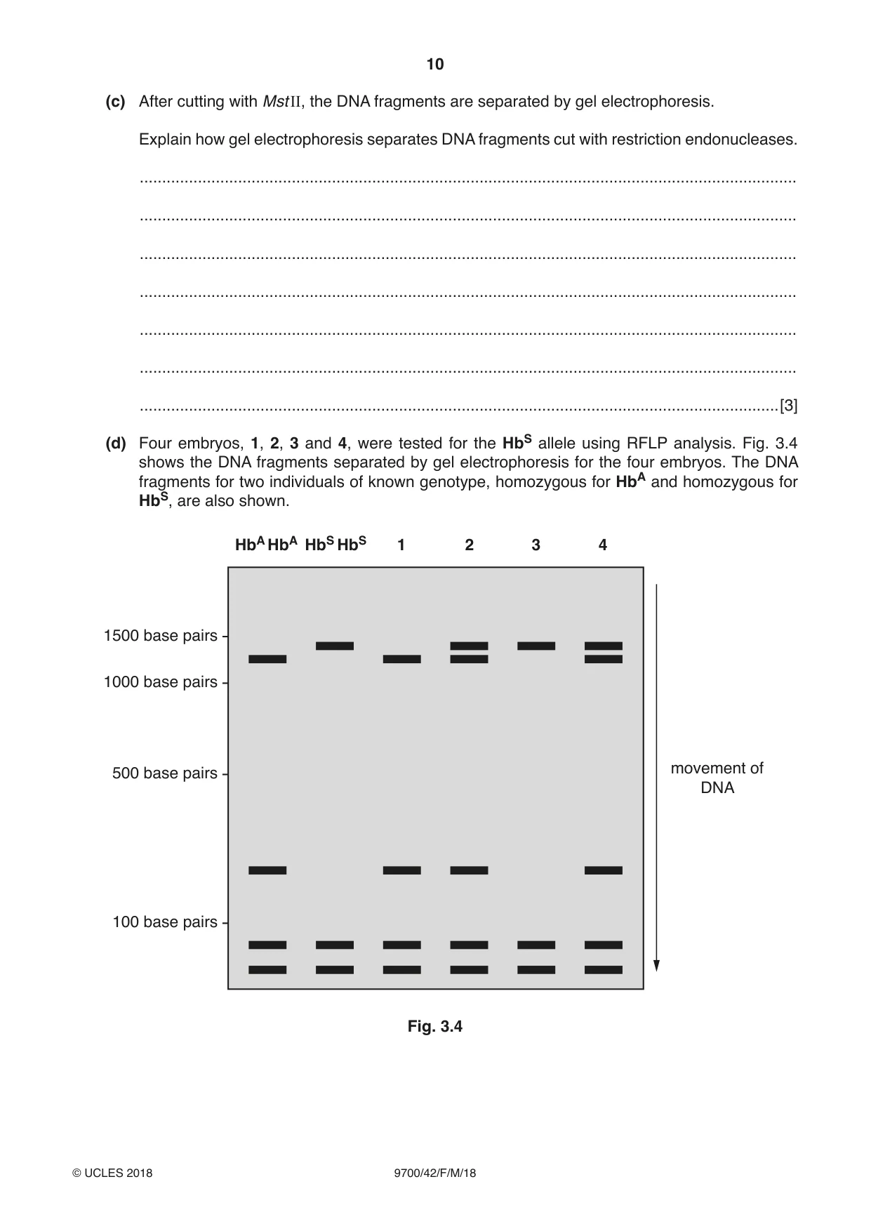
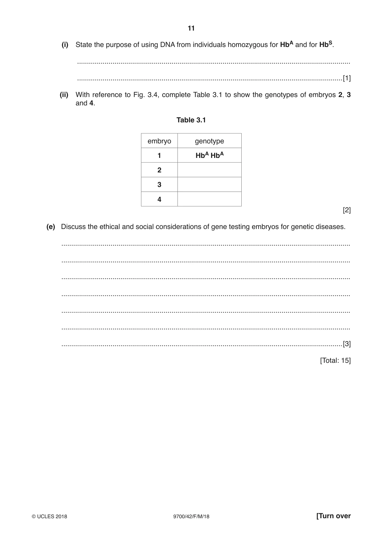
3 The β-globin gene codes for the β-globin polypeptide of haemoglobin. It has two alleles, HbA (normal) and HbS (sickle cell). The sickle cell allele differs from the normal allele due to a base substitution mutation and this mutation results in a single amino acid change to the β-globin polypeptide. There are three possible genotypes and phenotypes. • HbS HbS, sickle cell anaemia, a severe disease • HbA HbS, sickle cell trait with mild or no symptoms of sickle cell anaemia • HbA HbA, normal (healthy) A man and woman who both have sickle cell trait may choose to have children by IVF. This allows the genotype of embryos to be determined by gene testing before the embryos are implanted. Embryos with the normal genotype can then be selected and implanted into the mother. One technique that can be used in gene testing an embryo for the HbS allele is restriction fragment length polymorphism (RFLP) analysis. This involves digesting a DNA sample from an embryo with a restriction endonuclease and then separating the DNA fragments by gel electrophoresis. The position of the DNA fragments on the gel can show if the embryo has the HbS allele. (a) The first step in testing an embryo for the HbS allele by RFLP analysis requires many copies of the part of the β-globin gene in which the mutation causing sickle cell anaemia occurs. (i) Name the technique used to produce many copies of a DNA sequence from a very small quantity of DNA. .......................................................................................................................................[1] (ii) Explain why it is necessary to copy this DNA sequence many times in order to test embryos for HbS alleles by RFLP analysis. ........................................................................................................................................... .......................................................................................................................................[1] In the next step of RFLP analysis, the copies of the part of the β-globin gene from the first step are incubated with a restriction endonuclease, Mst II. This enzyme cuts at a specific sequence of DNA (the restriction site). The restriction site for Mst II is shown in Fig. 3.1. 5’ – C C T N A G G – 3’ 3’ – G G A N T C C – 5’ N = any nucleotide (A, T, C or G) Fig. 3.1 Fig. 3.2 shows the part of an HbA allele obtained from the first step. All the Mst II restriction sites and the number of DNA base pairs separating these restriction sites are shown. HbA 5’ – C C T T A G G C C T G A G G C C T T A G G – 3’ 3’ – G G A A T C C G G A C T C C G G A A T C C – 5’ 80 1200 200 90 base pairs base pairs base pairs base pairs Fig. 3.2 Fig. 3.3 shows the same part of an HbS allele. The single base substitution in the HbS allele that causes sickle cell anaemia is indicated. HbS 5’ – C C T T A G G C C T G T G G C C T T A G G – 3’ 3’ – G G A A T C C G G A C A C C G G A A T C C – 5’ 80 1200 200 90 base pairs base pairs base pairs base pairs single base pair substitution Fig. 3.3 (b) With reference to Fig. 3.1, Fig. 3.2 and Fig. 3.3, explain why the enzyme Mst II can be used in RFLP analysis to show the difference between these parts of the HbA and HbS alleles. ................................................................................................................................................... ................................................................................................................................................... ................................................................................................................................................... ................................................................................................................................................... ................................................................................................................................................... ................................................................................................................................................... ................................................................................................................................................... ................................................................................................................................................... (c) After cutting with Mst II, the DNA fragments are separated by gel electrophoresis. Explain how gel electrophoresis separates DNA fragments cut with restriction endonucleases. ................................................................................................................................................... ................................................................................................................................................... ................................................................................................................................................... ................................................................................................................................................... ................................................................................................................................................... ................................................................................................................................................... ...............................................................................................................................................[3] (d) Four embryos, 1, 2, 3 and 4, were tested for the HbS allele using RFLP analysis. Fig. 3.4 shows the DNA fragments separated by gel electrophoresis for the four embryos. The DNA fragments for two individuals of known genotype, homozygous for HbA and homozygous for HbS, are also shown. HbA HbA HbS HbS 1 2 3 4 1500 base pairs 1000 base pairs movement of 500 base pairs DNA 100 base pairs Fig. 3.4 (i) State the purpose of using DNA from individuals homozygous for HbA and for HbS. ........................................................................................................................................... .......................................................................................................................................[1] (ii) With reference to Fig. 3.4, complete Table 3.1 to show the genotypes of embryos 2, 3 and 4. Table 3.1 embryo genotype 1 HbA HbA 2 3 4 [2] (e) Discuss the ethical and social considerations of gene testing embryos for genetic diseases. ................................................................................................................................................... ................................................................................................................................................... ................................................................................................................................................... ................................................................................................................................................... ................................................................................................................................................... ................................................................................................................................................... ...............................................................................................................................................[3] [Total: 15]
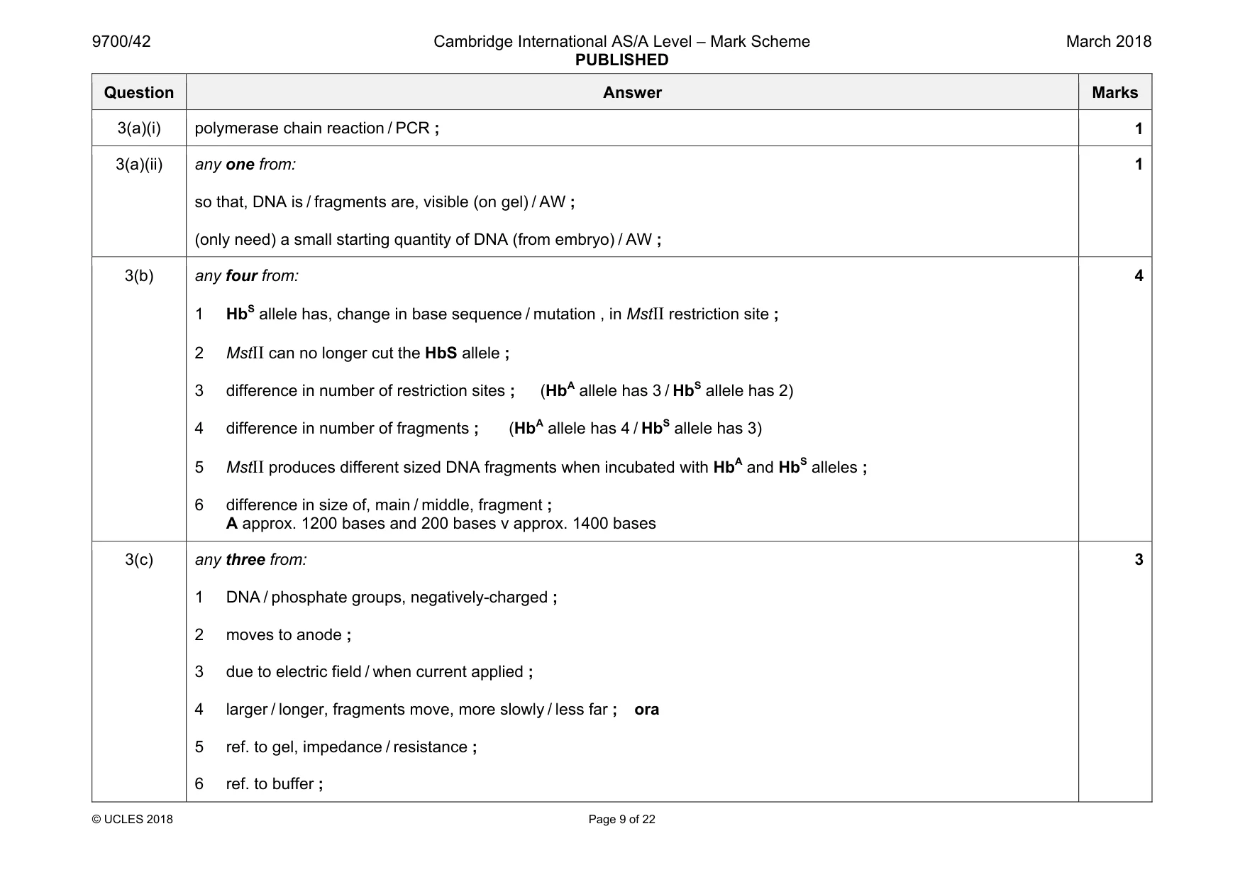
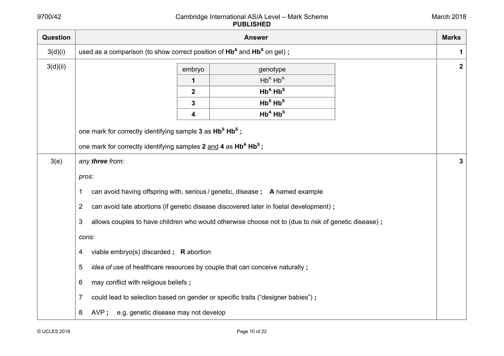
Mark scheme: 3(a)(i) polymerase chain reaction / PCR ; 1 3(a)(ii) any one from: so that, DNA is / fragments are, visible (on gel) / AW ; (only need) a small starting quantity of DNA (from embryo) / AW ; 1 3(b) any four from: 1 HbS allele has, change in base sequence / mutation , in MstII restriction site ; 2 MstII can no longer cut the HbS allele ; 3 difference in number of restriction sites ; (HbA allele has 3 / HbS allele has 2) 4 difference in number of fragments ; (HbA allele has 4 / HbS allele has 3) 5 MstII produces different sized DNA fragments when incubated with HbA and HbS alleles ; 6 difference in size of, main / middle, fragment ; A approx. 1200 bases and 200 bases v approx. 1400 bases 4 3(c) any three from: 1 DNA / phosphate groups, negatively-charged ; 2 moves to anode ; 3 due to electric field / when current applied ; 4 larger / longer, fragments move, more slowly / less far ; ora 5 ref. to gel, impedance / resistance ; 6 ref. to buffer ; 3 Question Answer Marks 3(d)(i) used as a comparison (to show correct position of HbA and HbS on gel) ; 1 3(d)(ii) embryo genotype 1 HbA HbA 2 HbA HbS 3 HbS HbS 4 HbA HbS one mark for correctly identifying sample 3 as HbS HbS ; one mark for correctly identifying samples 2 and 4 as HbA HbS ; 2 3(e) any three from: pros: 1 can avoid having offspring with, serious / genetic, disease ; A named example 2 can avoid late abortions (if genetic disease discovered later in foetal development) ; 3 allows couples to have children who would otherwise choose not to (due to risk of genetic disease) ; cons: 4 viable embryo(s) discarded ; R abortion 5 idea of use of healthcare resources by couple that can conceive naturally ; 6 may conflict with religious beliefs ; 7 could lead to selection based on gender or specific traits (“designer babies”) ; 8 AVP ; e.g. genetic disease may not develop 3
Q4 · Ribulose 1,5-bisphosphate carboxylase/oxygenase (rubisco) is an important enzyme involved…
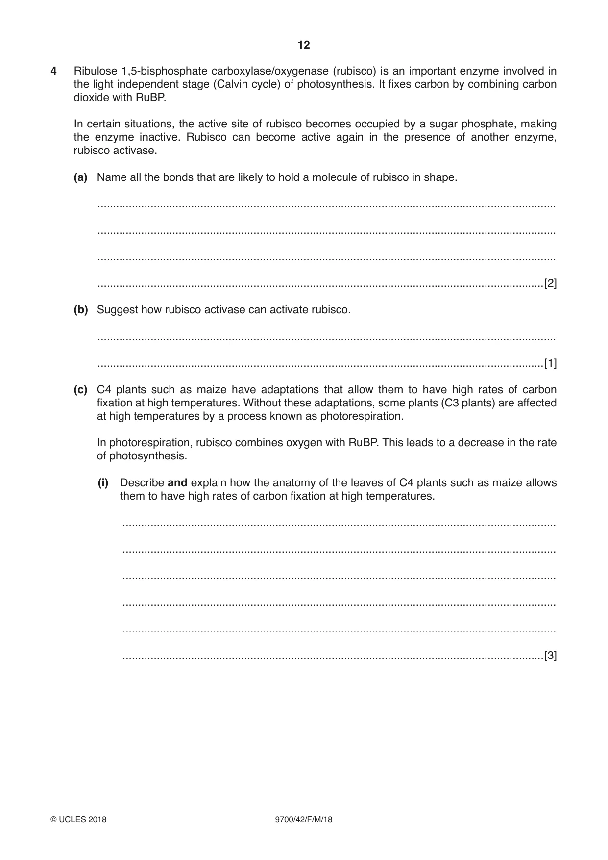
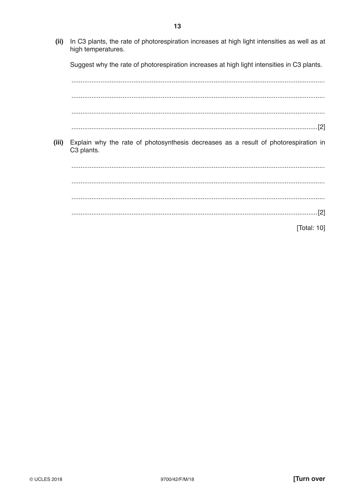
4 Ribulose 1,5-bisphosphate carboxylase/oxygenase (rubisco) is an important enzyme involved in the light independent stage (Calvin cycle) of photosynthesis. It fixes carbon by combining carbon dioxide with RuBP. In certain situations, the active site of rubisco becomes occupied by a sugar phosphate, making the enzyme inactive. Rubisco can become active again in the presence of another enzyme, rubisco activase. (a) Name all the bonds that are likely to hold a molecule of rubisco in shape. ................................................................................................................................................... ................................................................................................................................................... ................................................................................................................................................... ...............................................................................................................................................[2] (b) Suggest how rubisco activase can activate rubisco. ................................................................................................................................................... ...............................................................................................................................................[1] (c) C4 plants such as maize have adaptations that allow them to have high rates of carbon fixation at high temperatures. Without these adaptations, some plants (C3 plants) are affected at high temperatures by a process known as photorespiration. In photorespiration, rubisco combines oxygen with RuBP. This leads to a decrease in the rate of photosynthesis. (i) Describe and explain how the anatomy of the leaves of C4 plants such as maize allows them to have high rates of carbon fixation at high temperatures. ........................................................................................................................................... ........................................................................................................................................... ........................................................................................................................................... ........................................................................................................................................... ........................................................................................................................................... .......................................................................................................................................[3] (ii) In C3 plants, the rate of photorespiration increases at high light intensities as well as at high temperatures. Suggest why the rate of photorespiration increases at high light intensities in C3 plants. ........................................................................................................................................... ........................................................................................................................................... ........................................................................................................................................... .......................................................................................................................................[2] (iii) Explain why the rate of photosynthesis decreases as a result of photorespiration in C3 plants. ........................................................................................................................................... ........................................................................................................................................... ........................................................................................................................................... .......................................................................................................................................[2] [Total: 10]
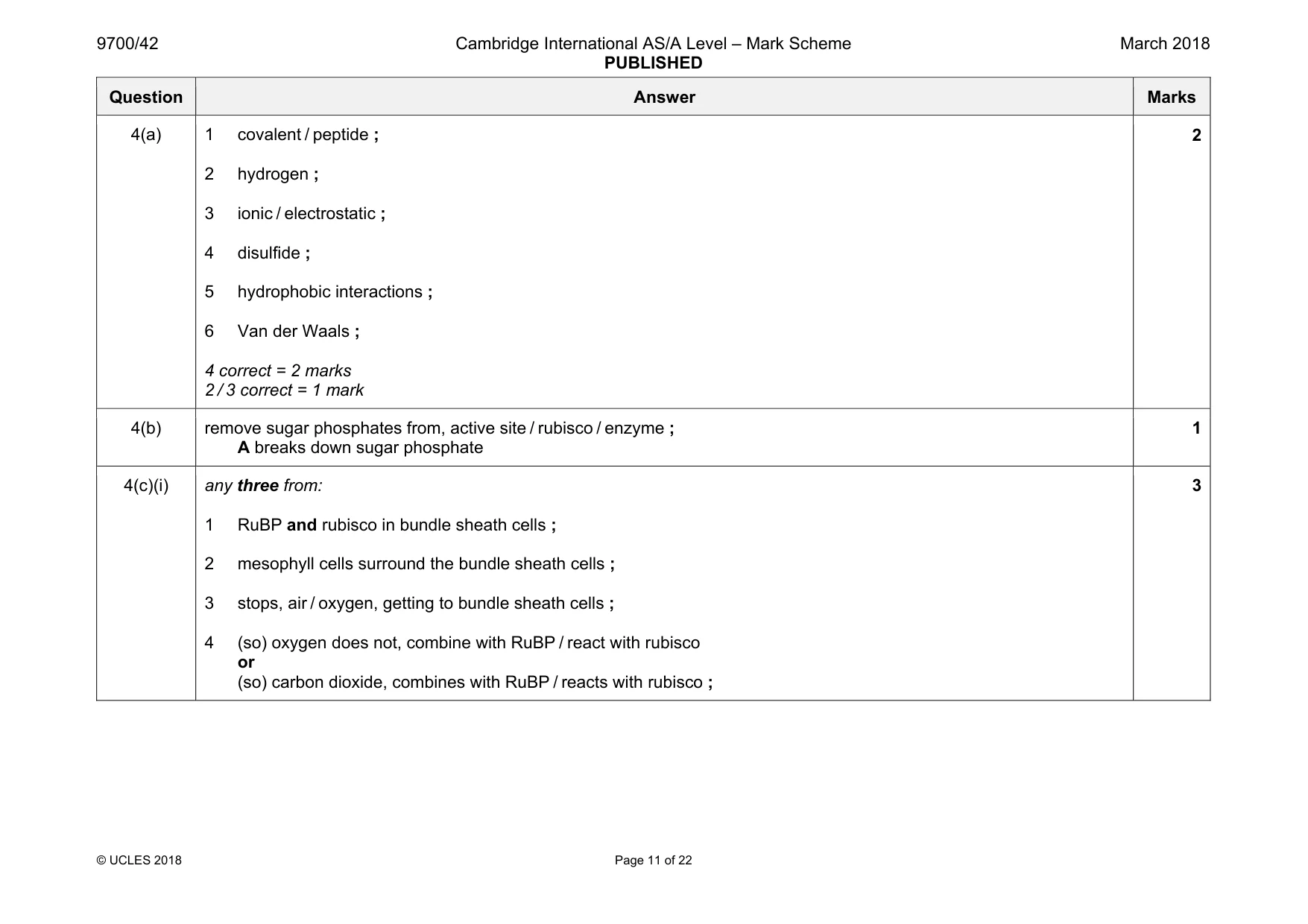
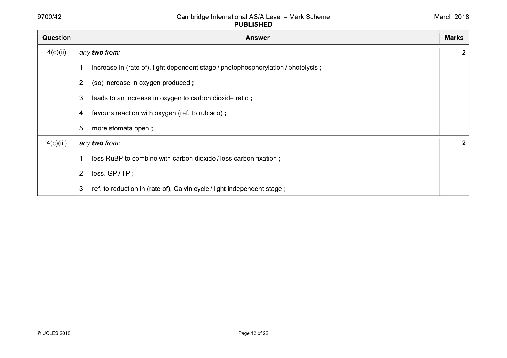
Mark scheme: 4(a) 1 covalent / peptide ; 2 hydrogen ; 3 ionic / electrostatic ; 4 disulfide ; 5 hydrophobic interactions ; 6 Van der Waals ; 4 correct = 2 marks 2 / 3 correct = 1 mark 2 4(b) remove sugar phosphates from, active site / rubisco / enzyme ; A breaks down sugar phosphate 1 4(c)(i) any three from: 1 RuBP and rubisco in bundle sheath cells ; 2 mesophyll cells surround the bundle sheath cells ; 3 stops, air / oxygen, getting to bundle sheath cells ; 4 (so) oxygen does not, combine with RuBP / react with rubisco or (so) carbon dioxide, combines with RuBP / reacts with rubisco ; 3 Question Answer Marks 4(c)(ii) any two from: 1 increase in (rate of), light dependent stage / photophosphorylation / photolysis ; 2 (so) increase in oxygen produced ; 3 leads to an increase in oxygen to carbon dioxide ratio ; 4 favours reaction with oxygen (ref. to rubisco) ; 5 more stomata open ; 2 4(c)(iii) any two from: 1 less RuBP to combine with carbon dioxide / less carbon fixation ; 2 less, GP / TP ; 3 ref. to reduction in (rate of), Calvin cycle / light independent stage ; 2
More questions on Photosynthesis as an energy transfer process
Q5 · The contraction of striated muscle is explained by the sliding filament model
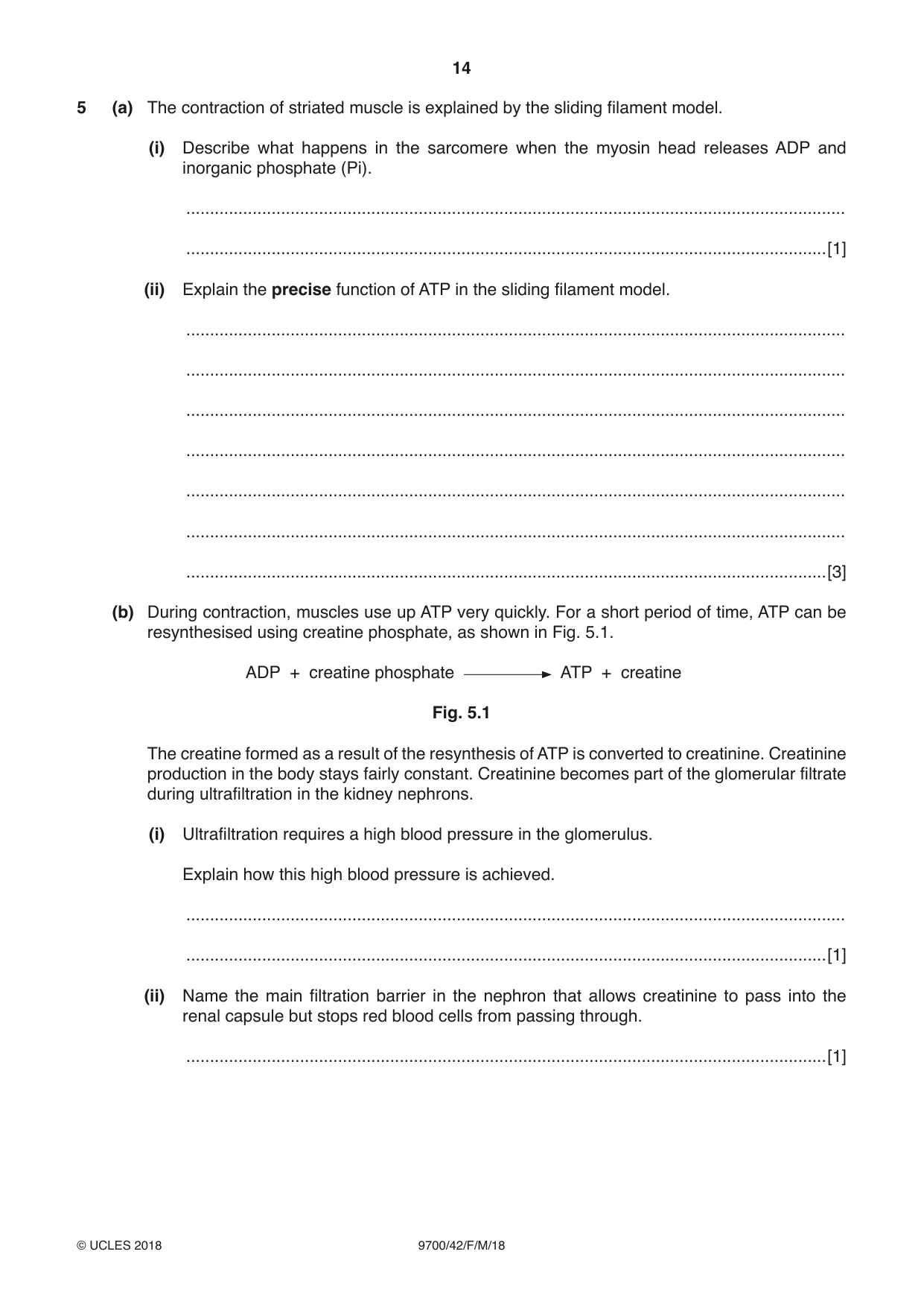
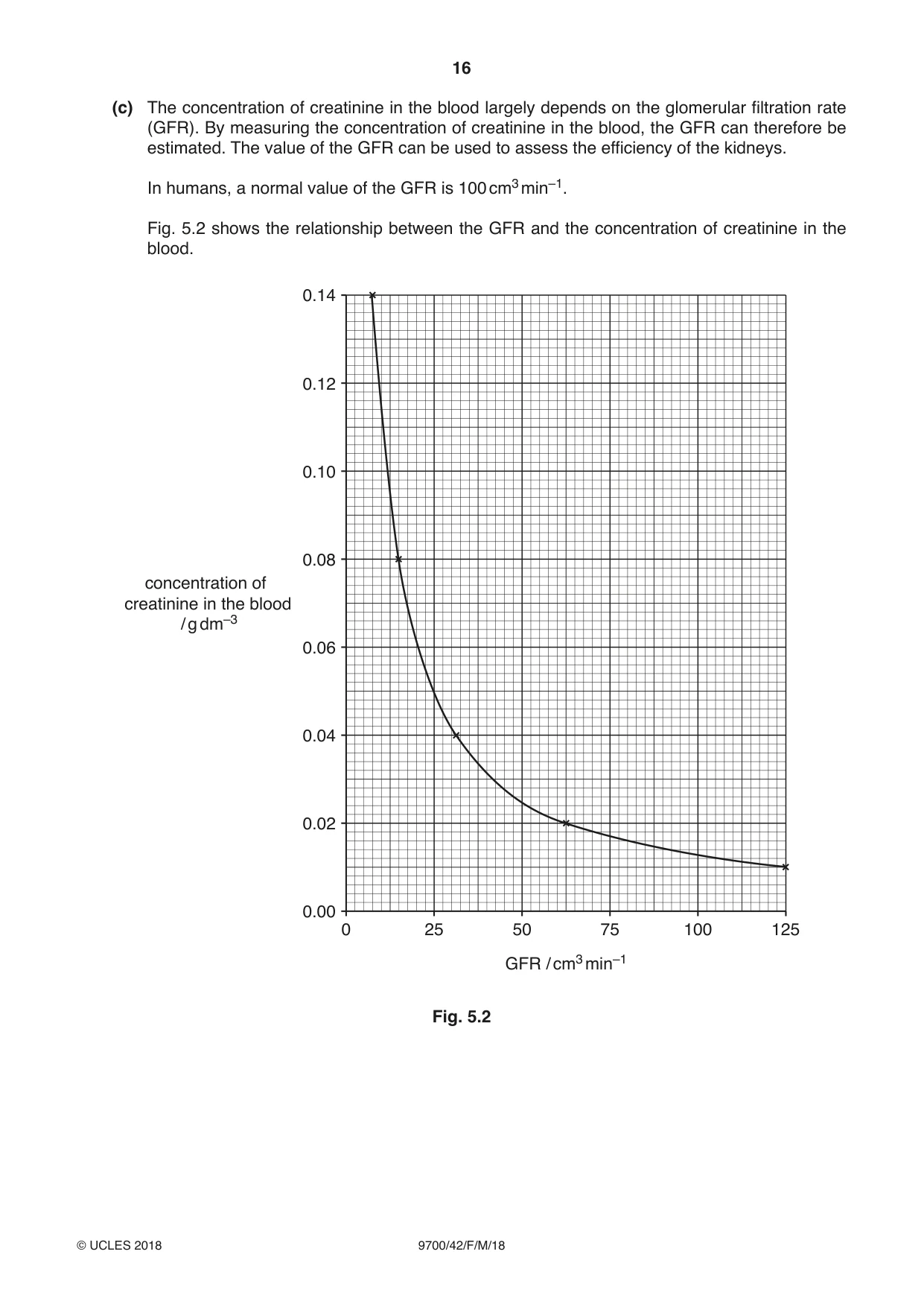
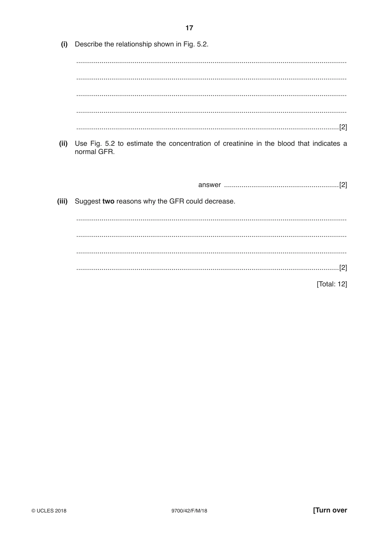
5 (a) The contraction of striated muscle is explained by the sliding filament model. (i) Describe what happens in the sarcomere when the myosin head releases ADP and inorganic phosphate (Pi). ........................................................................................................................................... .......................................................................................................................................[1] (ii) Explain the precise function of ATP in the sliding filament model. ........................................................................................................................................... ........................................................................................................................................... ........................................................................................................................................... ........................................................................................................................................... ........................................................................................................................................... ........................................................................................................................................... .......................................................................................................................................[3] (b) During contraction, muscles use up ATP very quickly. For a short period of time, ATP can be resynthesised using creatine phosphate, as shown in Fig. 5.1. ADP + creatine phosphate ATP + creatine Fig. 5.1 The creatine formed as a result of the resynthesis of ATP is converted to creatinine. Creatinine production in the body stays fairly constant. Creatinine becomes part of the glomerular filtrate during ultrafiltration in the kidney nephrons. (i) Ultrafiltration requires a high blood pressure in the glomerulus. Explain how this high blood pressure is achieved. ........................................................................................................................................... .......................................................................................................................................[1] (ii) Name the main filtration barrier in the nephron that allows creatinine to pass into the renal capsule but stops red blood cells from passing through. .......................................................................................................................................[1] Question 5 continues on page 16 (c) The concentration of creatinine in the blood largely depends on the glomerular filtration rate (GFR). By measuring the concentration of creatinine in the blood, the GFR can therefore be estimated. The value of the GFR can be used to assess the efficiency of the kidneys. In humans, a normal value of the GFR is 100 cm3 min–1. Fig. 5.2 shows the relationship between the GFR and the concentration of creatinine in the blood. 0.14 0.12 0.10 0.08 concentration of creatinine in the blood / g dm–3 0.06 0.04 0.02 0.00 0 25 50 75 100 125 GFR / cm3 min–1 Fig. 5.2 (i) Describe the relationship shown in Fig. 5.2. ........................................................................................................................................... ........................................................................................................................................... ........................................................................................................................................... ........................................................................................................................................... .......................................................................................................................................[2] (ii) Use Fig. 5.2 to estimate the concentration of creatinine in the blood that indicates a normal GFR. answer ...........................................................[2] (iii) Suggest two reasons why the GFR could decrease. ........................................................................................................................................... ........................................................................................................................................... ........................................................................................................................................... .......................................................................................................................................[2] [Total: 12]
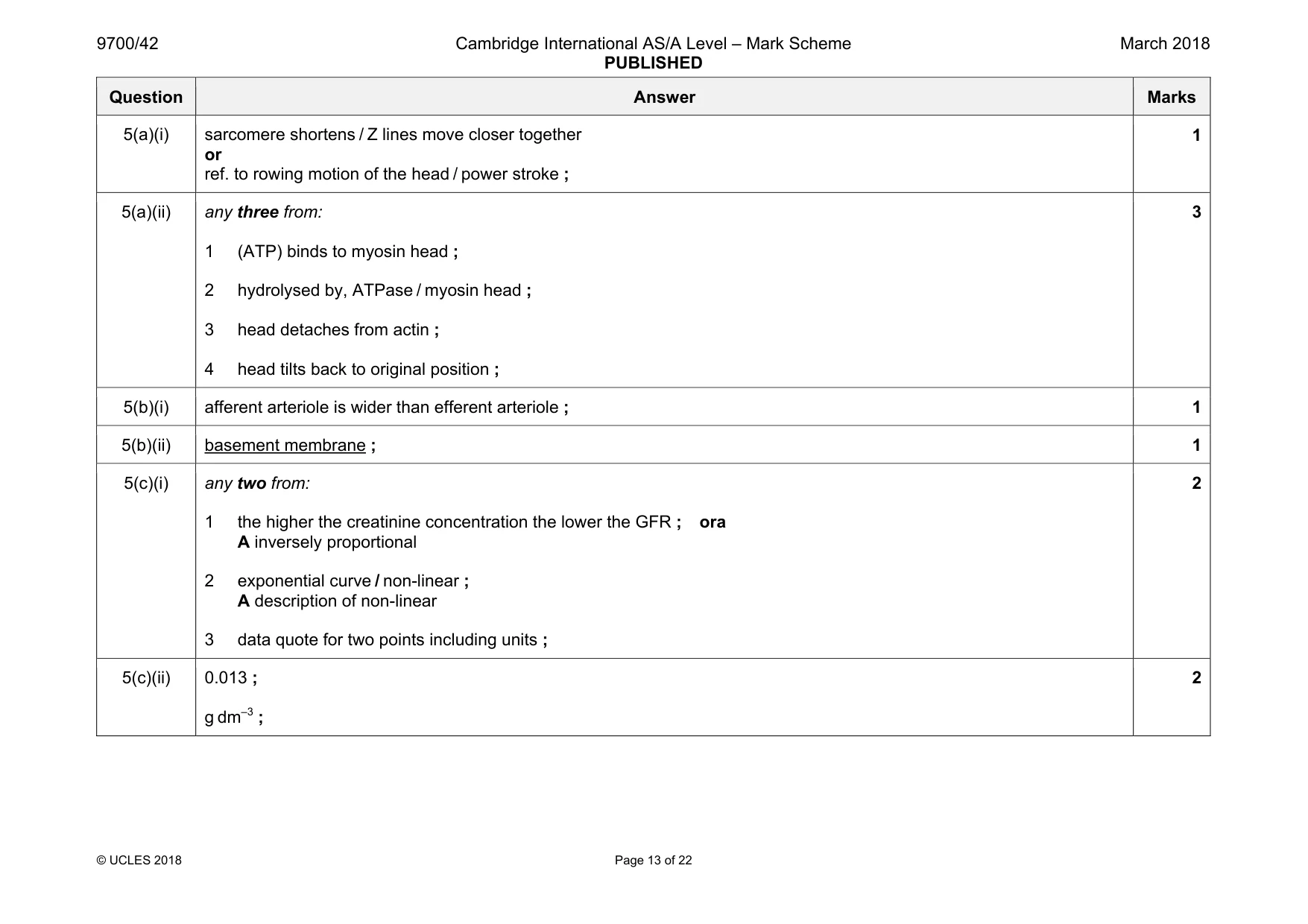
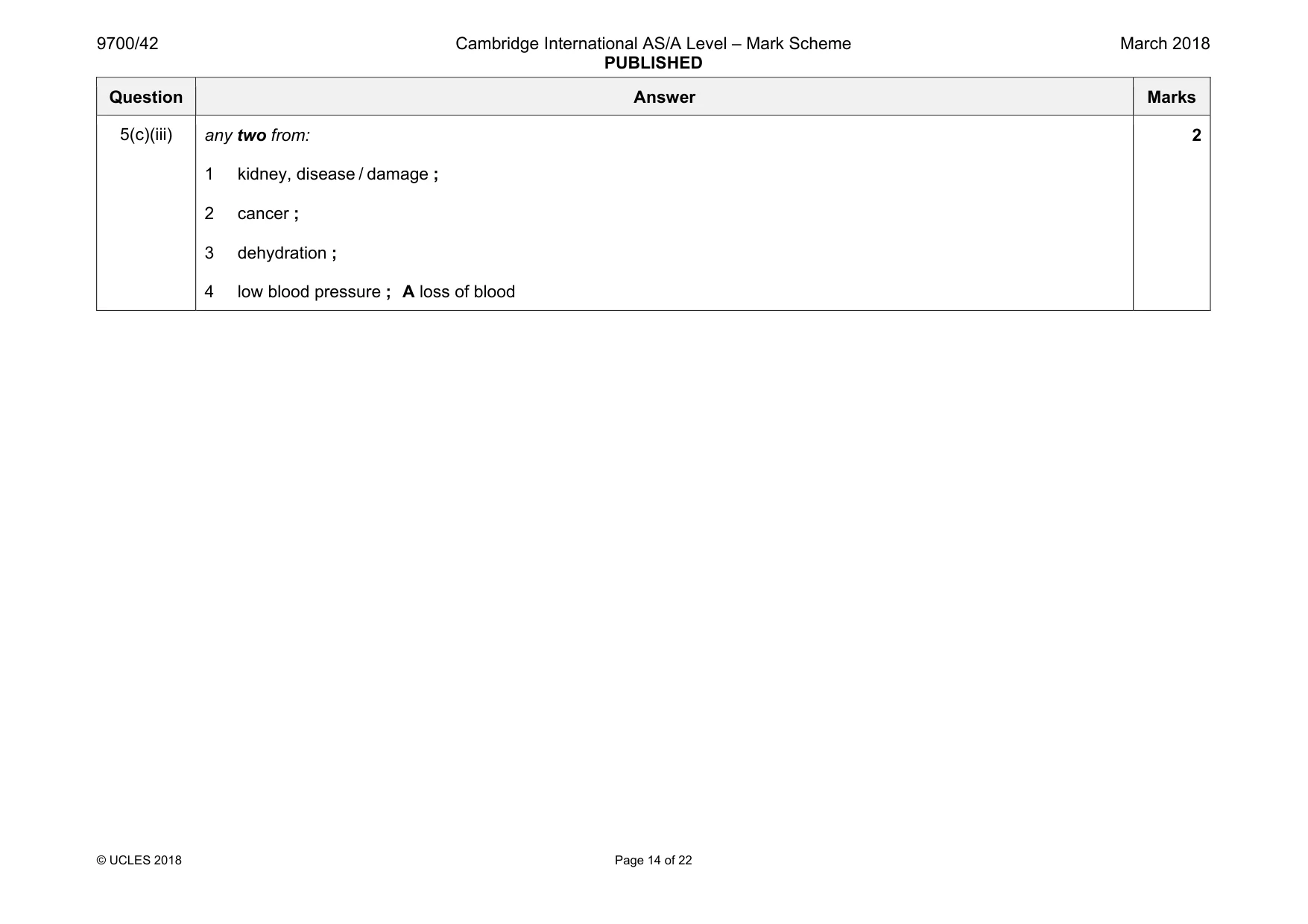
Mark scheme: 5(a)(i) sarcomere shortens / Z lines move closer together or ref. to rowing motion of the head / power stroke ; 1 5(a)(ii) any three from: 1 (ATP) binds to myosin head ; 2 hydrolysed by, ATPase / myosin head ; 3 head detaches from actin ; 4 head tilts back to original position ; 3 5(b)(i) afferent arteriole is wider than efferent arteriole ; 1 5(b)(ii) basement membrane ; 1 5(c)(i) any two from: 1 the higher the creatinine concentration the lower the GFR ; ora A inversely proportional 2 exponential curve / non-linear ; A description of non-linear 3 data quote for two points including units ; 2 5(c)(ii) 0.013 ; g dm–3 ; 2 Question Answer Marks 5(c)(iii) any two from: 1 kidney, disease / damage ; 2 cancer ; 3 dehydration ; 4 low blood pressure ; A loss of blood 2
Q6 · The black pigment melanin, which contributes to hair, skin and eye colour, is produced by…
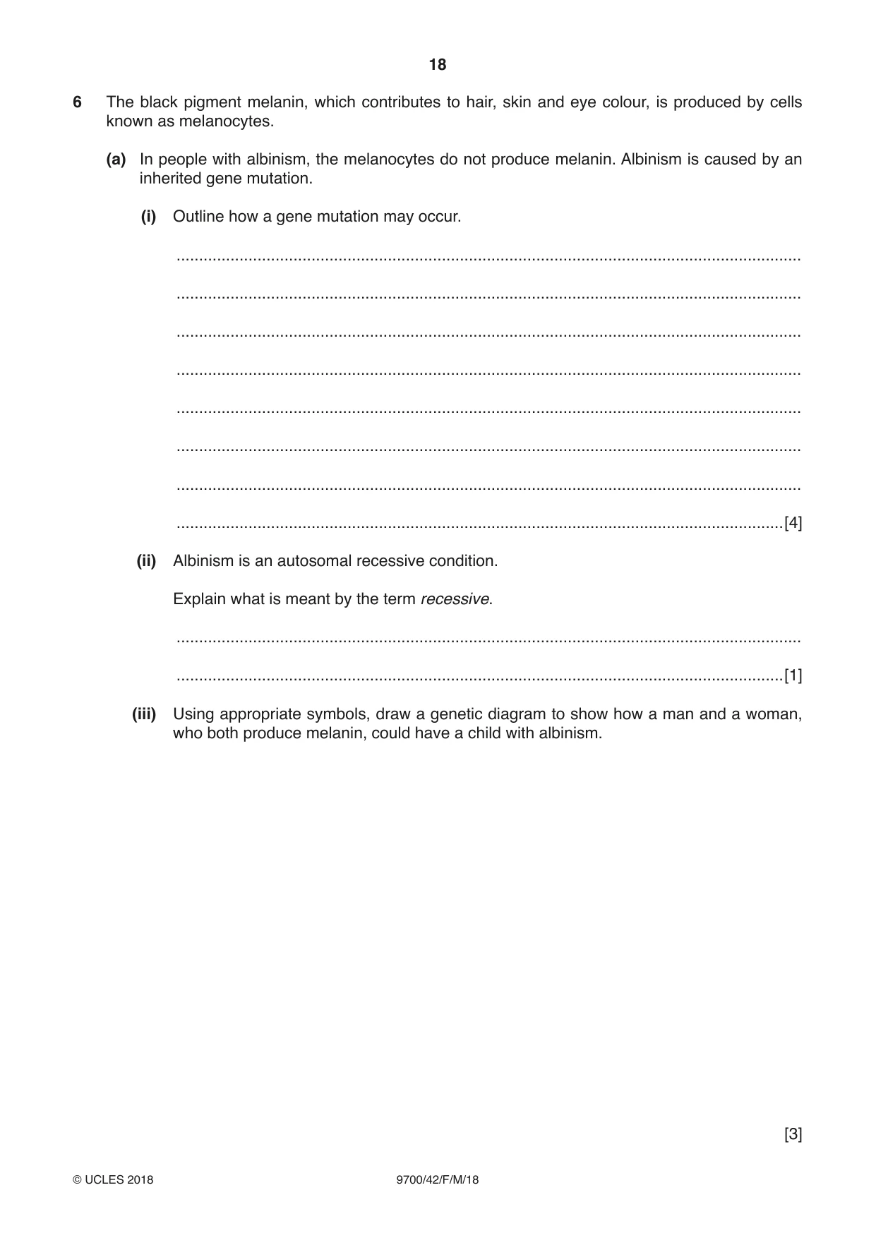
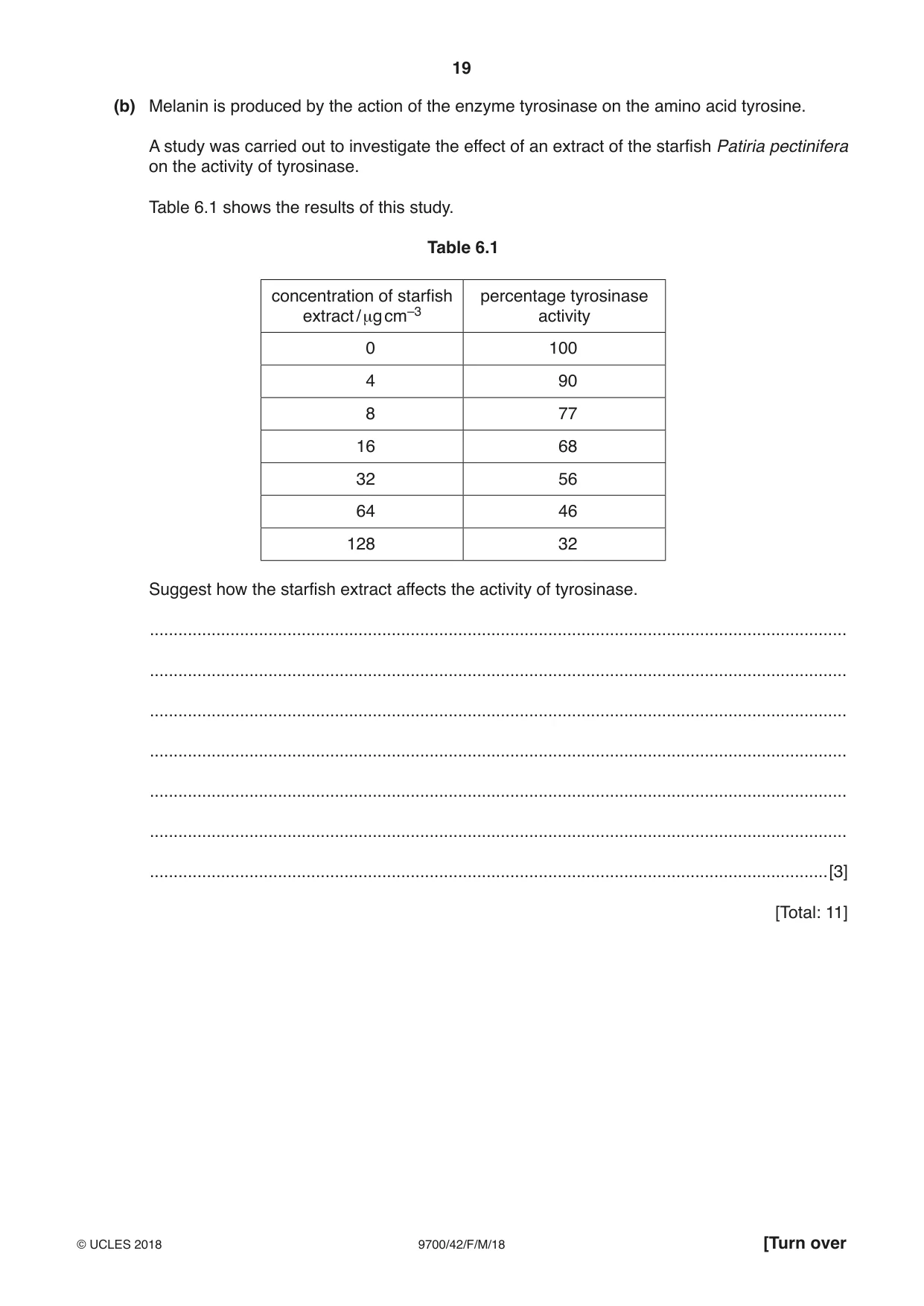
6 The black pigment melanin, which contributes to hair, skin and eye colour, is produced by cells known as melanocytes. (a) In people with albinism, the melanocytes do not produce melanin. Albinism is caused by an inherited gene mutation. (i) Outline how a gene mutation may occur. ........................................................................................................................................... ........................................................................................................................................... ........................................................................................................................................... ........................................................................................................................................... ........................................................................................................................................... ........................................................................................................................................... ........................................................................................................................................... .......................................................................................................................................[4] (ii) Albinism is an autosomal recessive condition. Explain what is meant by the term recessive. ........................................................................................................................................... .......................................................................................................................................[1] (iii) Using appropriate symbols, draw a genetic diagram to show how a man and a woman, who both produce melanin, could have a child with albinism. [3] (b) Melanin is produced by the action of the enzyme tyrosinase on the amino acid tyrosine. A study was carried out to investigate the effect of an extract of the starfish Patiria pectinifera on the activity of tyrosinase. Table 6.1 shows the results of this study. Table 6.1 concentration of starfish percentage tyrosinase extract / µg cm–3 activity 0 100 4 90 8 77 16 68 32 56 64 46 128 32 Suggest how the starfish extract affects the activity of tyrosinase. ................................................................................................................................................... ................................................................................................................................................... ................................................................................................................................................... ................................................................................................................................................... ................................................................................................................................................... ................................................................................................................................................... ...............................................................................................................................................[3] [Total: 11]
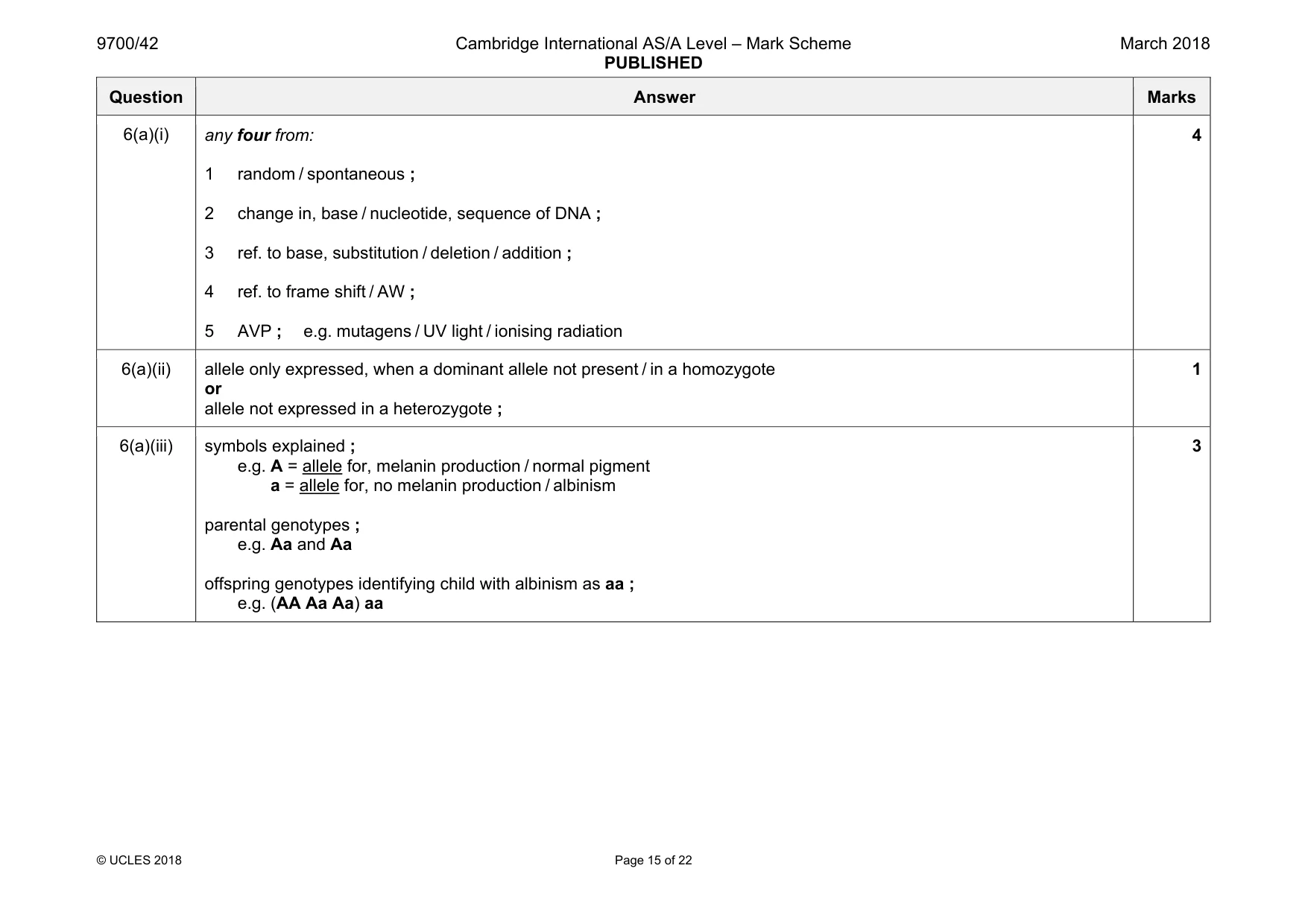
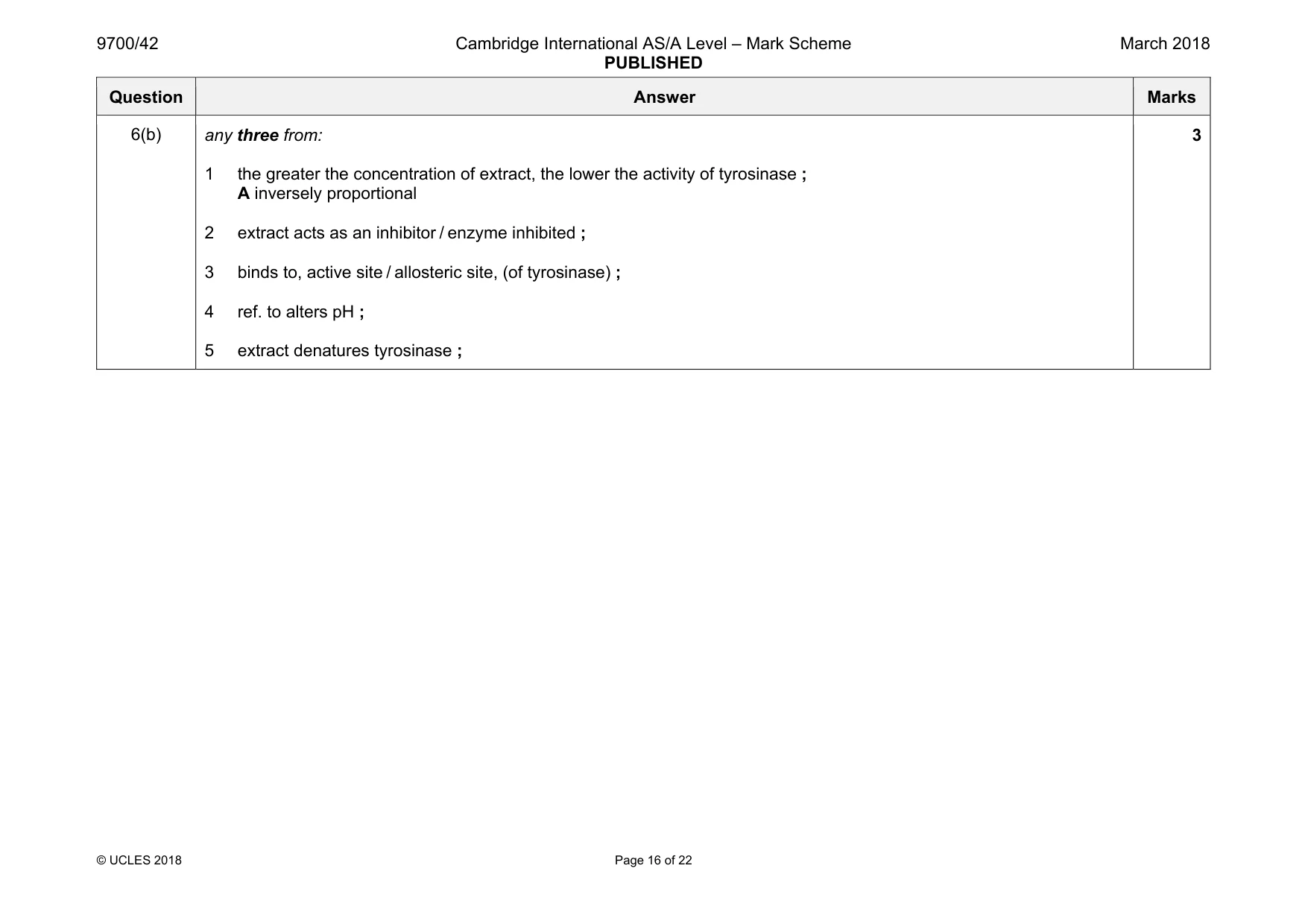
Mark scheme: 6(a)(i) any four from: 1 random / spontaneous ; 2 change in, base / nucleotide, sequence of DNA ; 3 ref. to base, substitution / deletion / addition ; 4 ref. to frame shift / AW ; 5 AVP ; e.g. mutagens / UV light / ionising radiation 4 6(a)(ii) allele only expressed, when a dominant allele not present / in a homozygote or allele not expressed in a heterozygote ; 1 6(a)(iii) symbols explained ; e.g. A = allele for, melanin production / normal pigment a = allele for, no melanin production / albinism parental genotypes ; e.g. Aa and Aa offspring genotypes identifying child with albinism as aa ; e.g. (AA Aa Aa) aa 3 Question Answer Marks 6(b) any three from: 1 the greater the concentration of extract, the lower the activity of tyrosinase ; A inversely proportional 2 extract acts as an inhibitor / enzyme inhibited ; 3 binds to, active site / allosteric site, (of tyrosinase) ; 4 ref. to alters pH ; 5 extract denatures tyrosinase ; 3
More questions on Passage of information from parents to offspring
Q7 · A red deer, Cervus elaphus
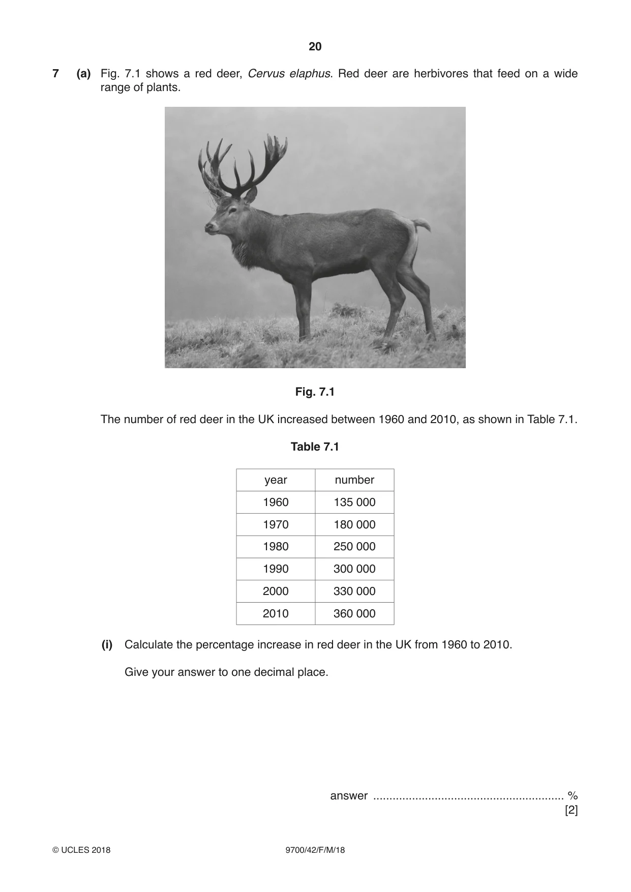
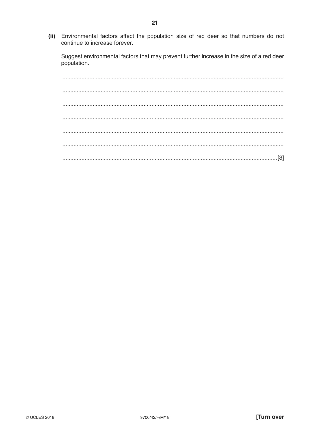
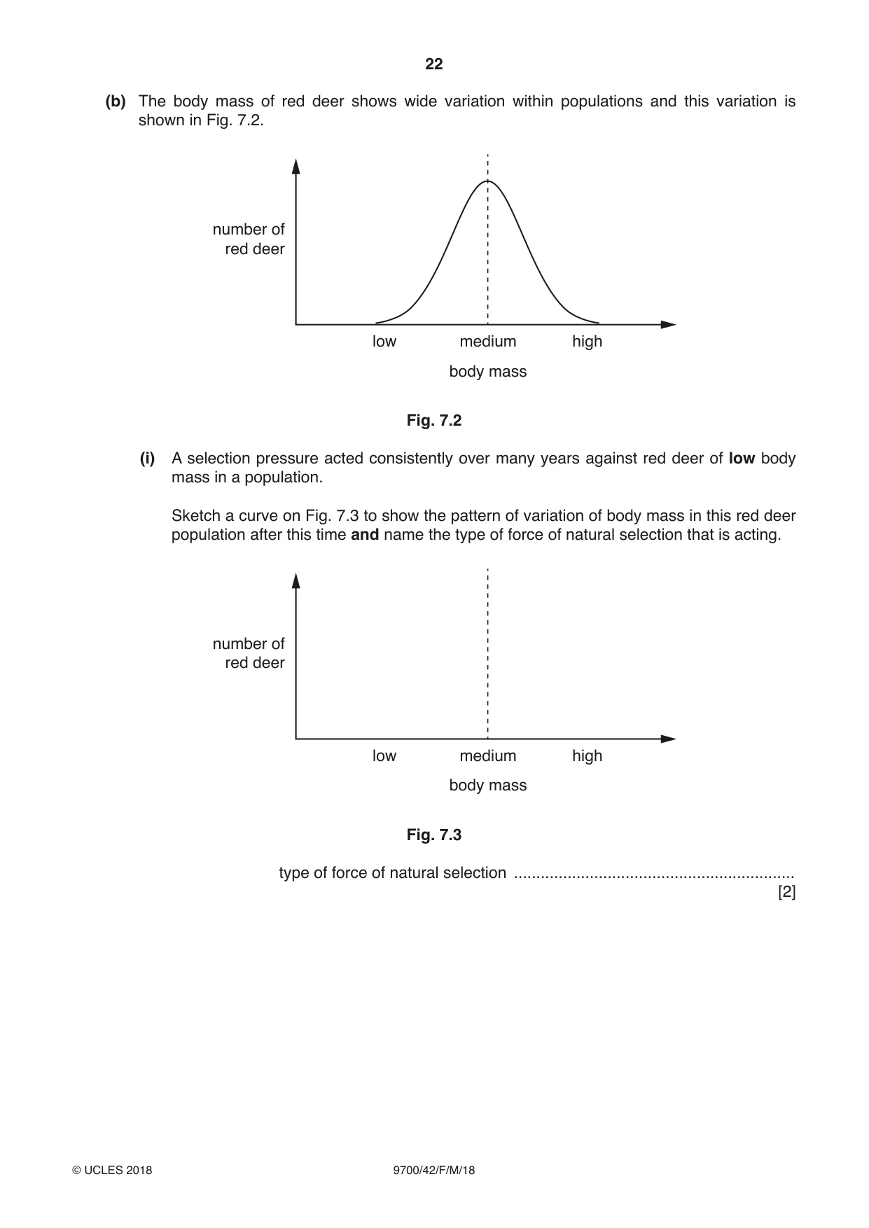
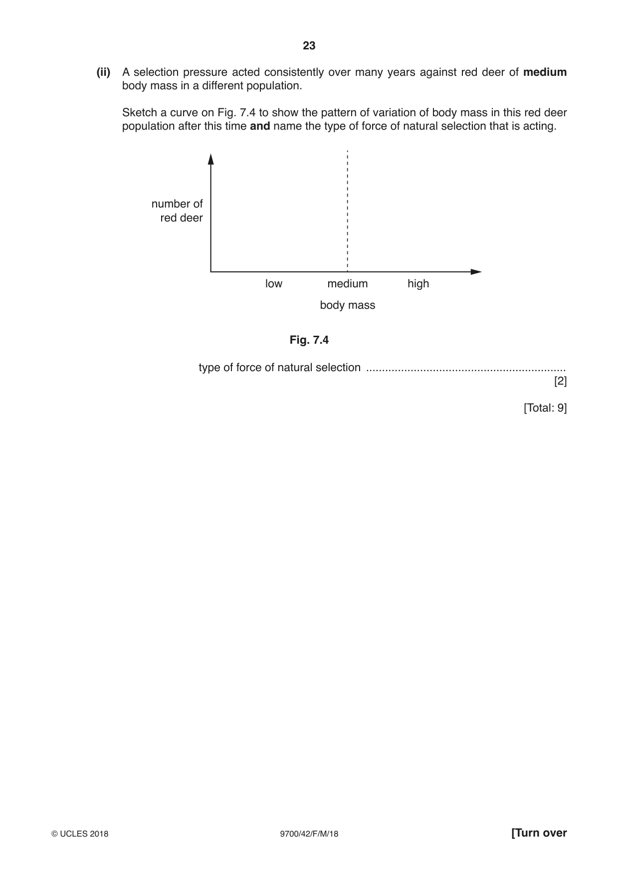
7 (a) Fig. 7.1 shows a red deer, Cervus elaphus. Red deer are herbivores that feed on a wide range of plants. Fig. 7.1 The number of red deer in the UK increased between 1960 and 2010, as shown in Table 7.1. Table 7.1 year number 1960 135 000 1970 180 000 1980 250 000 1990 300 000 2000 330 000 2010 360 000 (i) Calculate the percentage increase in red deer in the UK from 1960 to 2010. Give your answer to one decimal place. answer ........................................................... % [2] (ii) Environmental factors affect the population size of red deer so that numbers do not continue to increase forever. Suggest environmental factors that may prevent further increase in the size of a red deer population. ........................................................................................................................................... ........................................................................................................................................... ........................................................................................................................................... ........................................................................................................................................... ........................................................................................................................................... ........................................................................................................................................... .......................................................................................................................................[3] (b) The body mass of red deer shows wide variation within populations and this variation is shown in Fig. 7.2. number of red deer low medium high body mass Fig. 7.2 (i) A selection pressure acted consistently over many years against red deer of low body mass in a population. Sketch a curve on Fig. 7.3 to show the pattern of variation of body mass in this red deer population after this time and name the type of force of natural selection that is acting. number of red deer low medium high body mass Fig. 7.3 type of force of natural selection ............................................................... [2] (ii) A selection pressure acted consistently over many years against red deer of medium body mass in a different population. Sketch a curve on Fig. 7.4 to show the pattern of variation of body mass in this red deer population after this time and name the type of force of natural selection that is acting. number of red deer low medium high body mass Fig. 7.4 type of force of natural selection ............................................................... [2] [Total: 9]
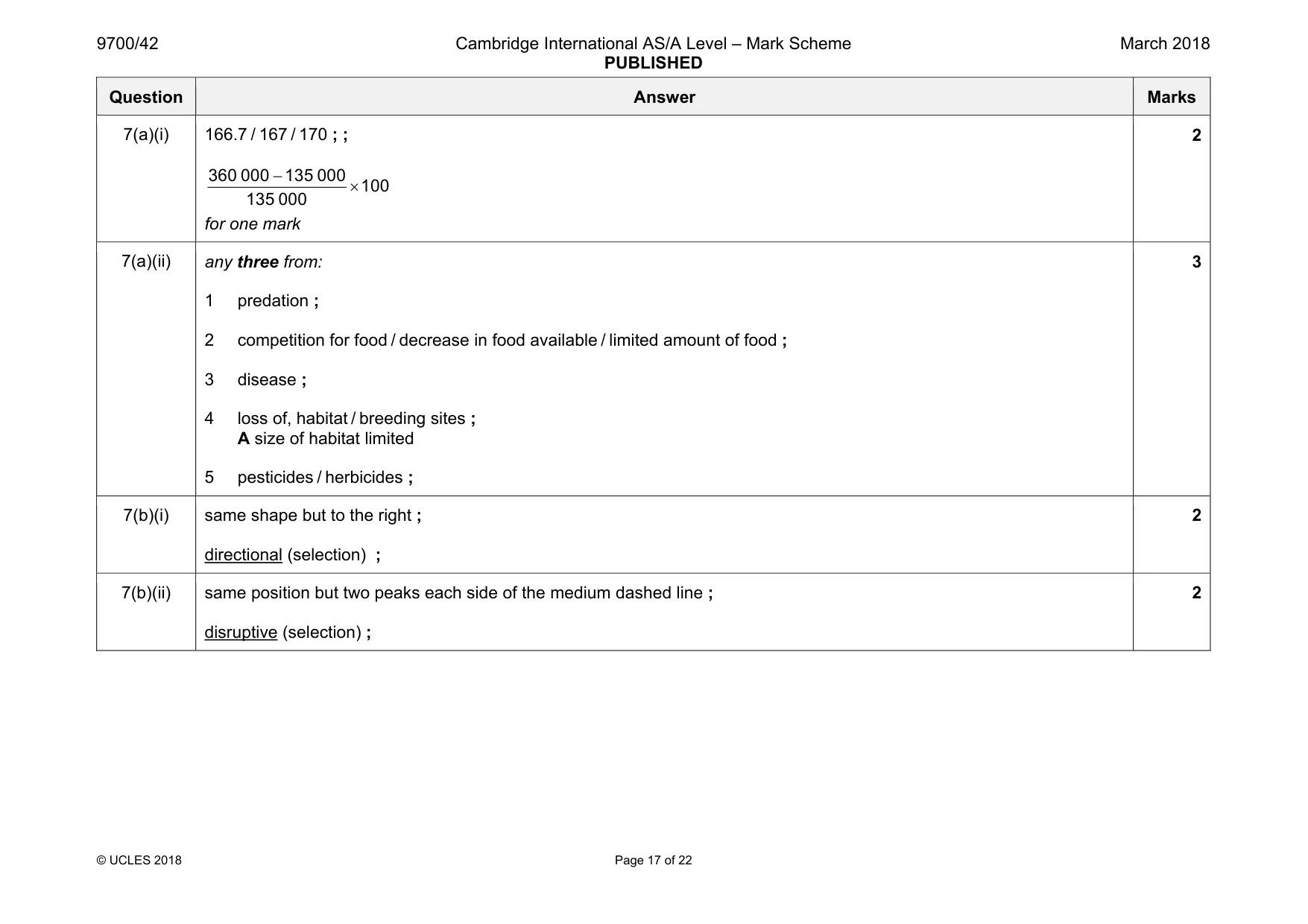
Mark scheme: 7(a)(i) 166.7 / 167 / 170 ; ; − × 360 000 135 000 100 135 000 for one mark 2 7(a)(ii) any three from: 1 predation ; 2 competition for food / decrease in food available / limited amount of food ; 3 disease ; 4 loss of, habitat / breeding sites ; A size of habitat limited 5 pesticides / herbicides ; 3 7(b)(i) same shape but to the right ; directional (selection) ; 2 7(b)(ii) same position but two peaks each side of the medium dashed line ; disruptive (selection) ; 2
Q8 · Structures and compounds involved in respiration include: 1 coenzyme A 2 cytoplasm 3…
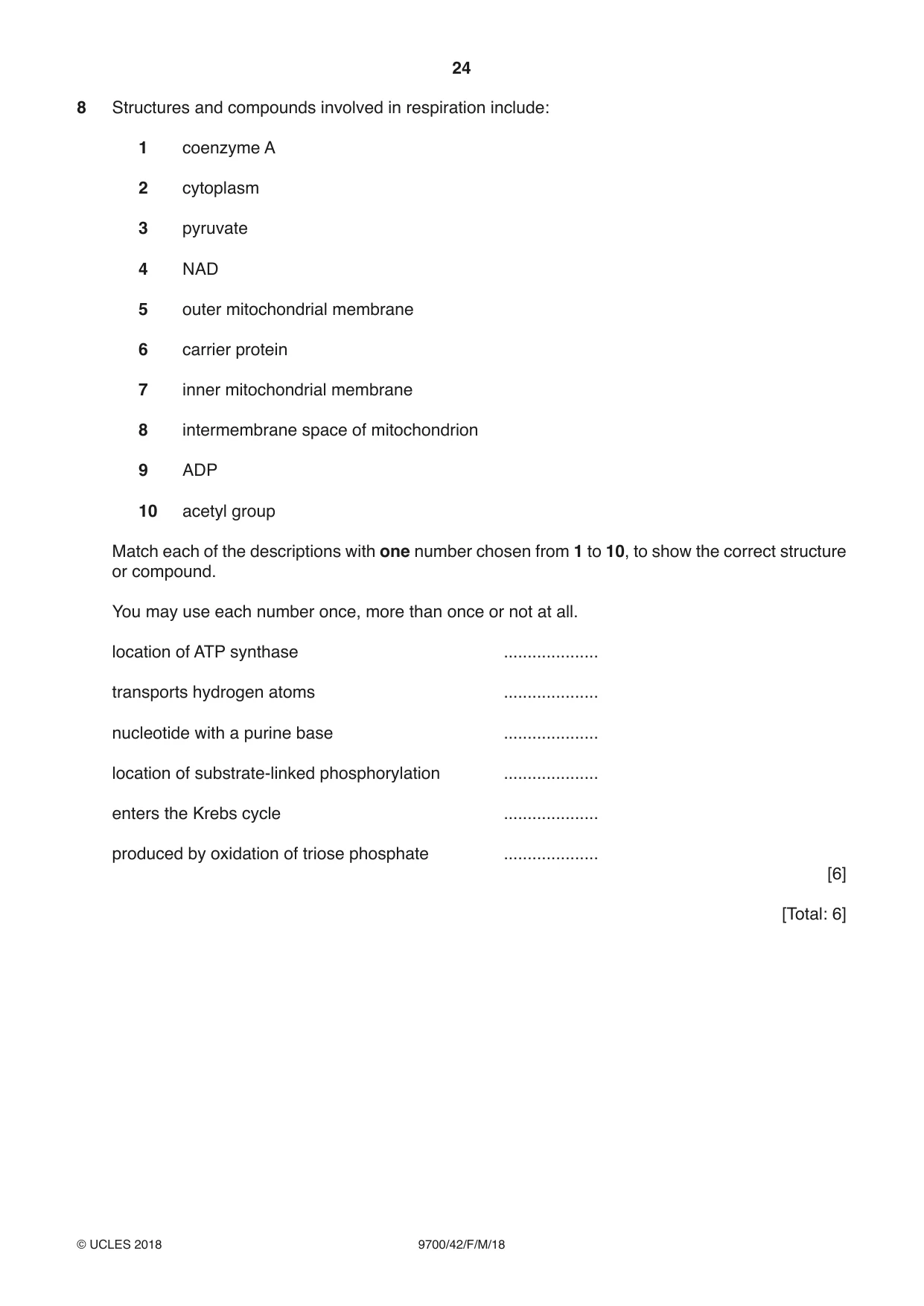
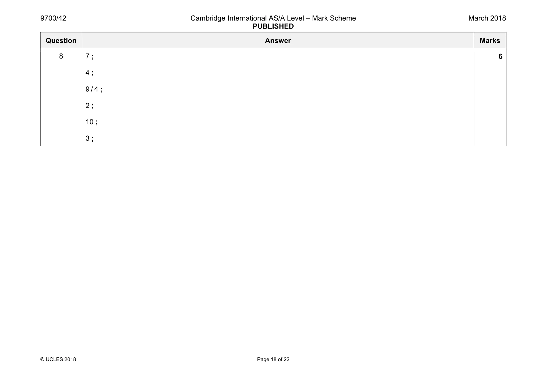
8 Structures and compounds involved in respiration include: 1 coenzyme A 2 cytoplasm 3 pyruvate 4 NAD 5 outer mitochondrial membrane 6 carrier protein 7 inner mitochondrial membrane 8 intermembrane space of mitochondrion 9 ADP 10 acetyl group Match each of the descriptions with one number chosen from 1 to 10, to show the correct structure or compound. You may use each number once, more than once or not at all. location of ATP synthase .................... transports hydrogen atoms .................... nucleotide with a purine base .................... location of substrate-linked phosphorylation .................... enters the Krebs cycle .................... produced by oxidation of triose phosphate .................... [6] [Total: 6]
Mark scheme: 8 7 ; 4 ; 9 / 4 ; 2 ; 10 ; 3 ; 6
Q9 · Describe the process of cyclic photophosphorylation and the structure of the photosystem…
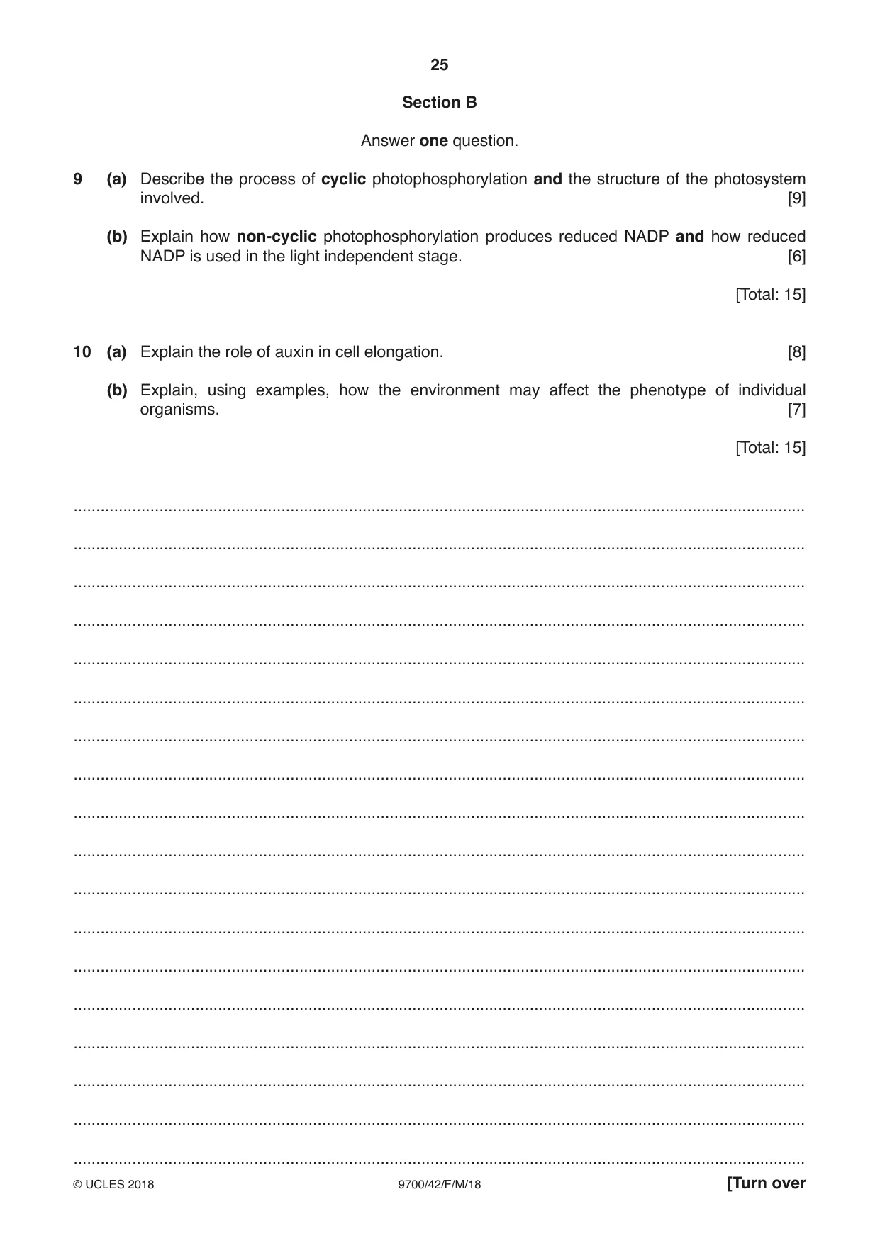
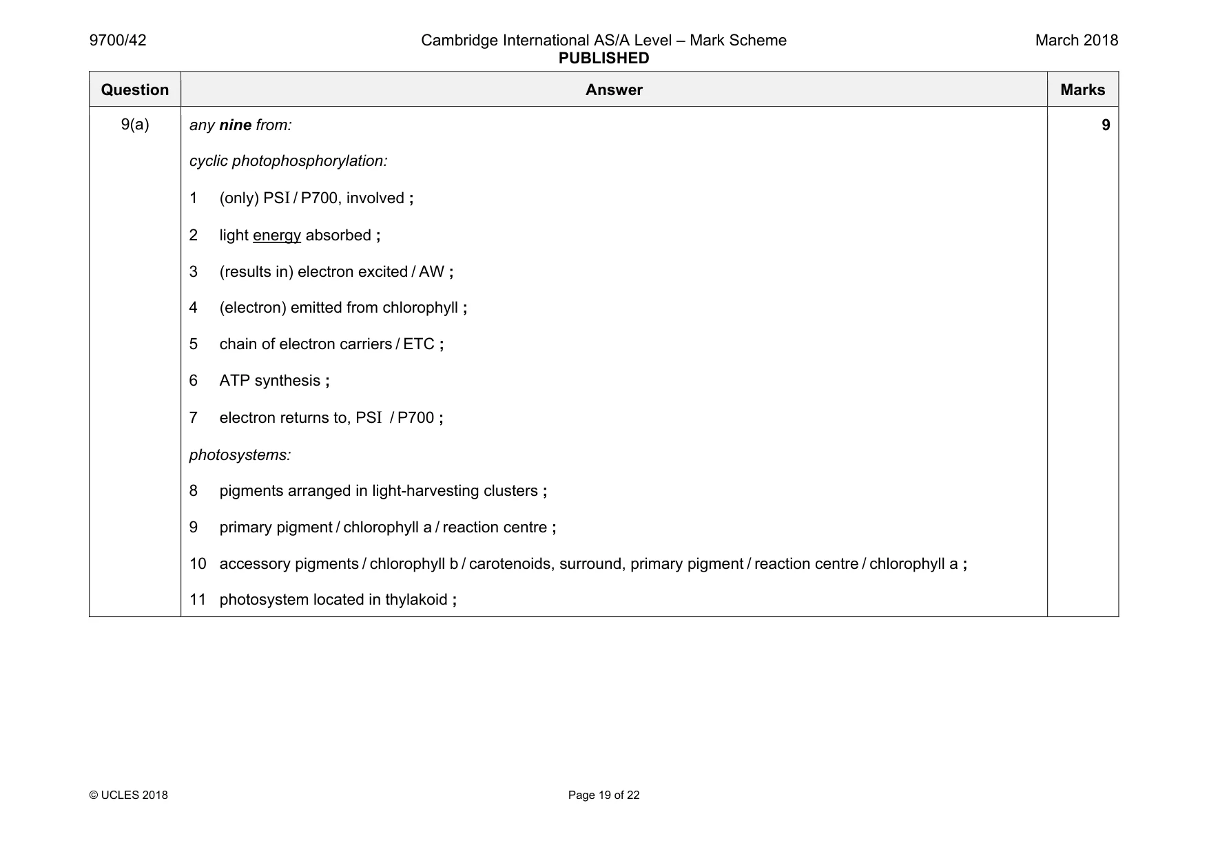
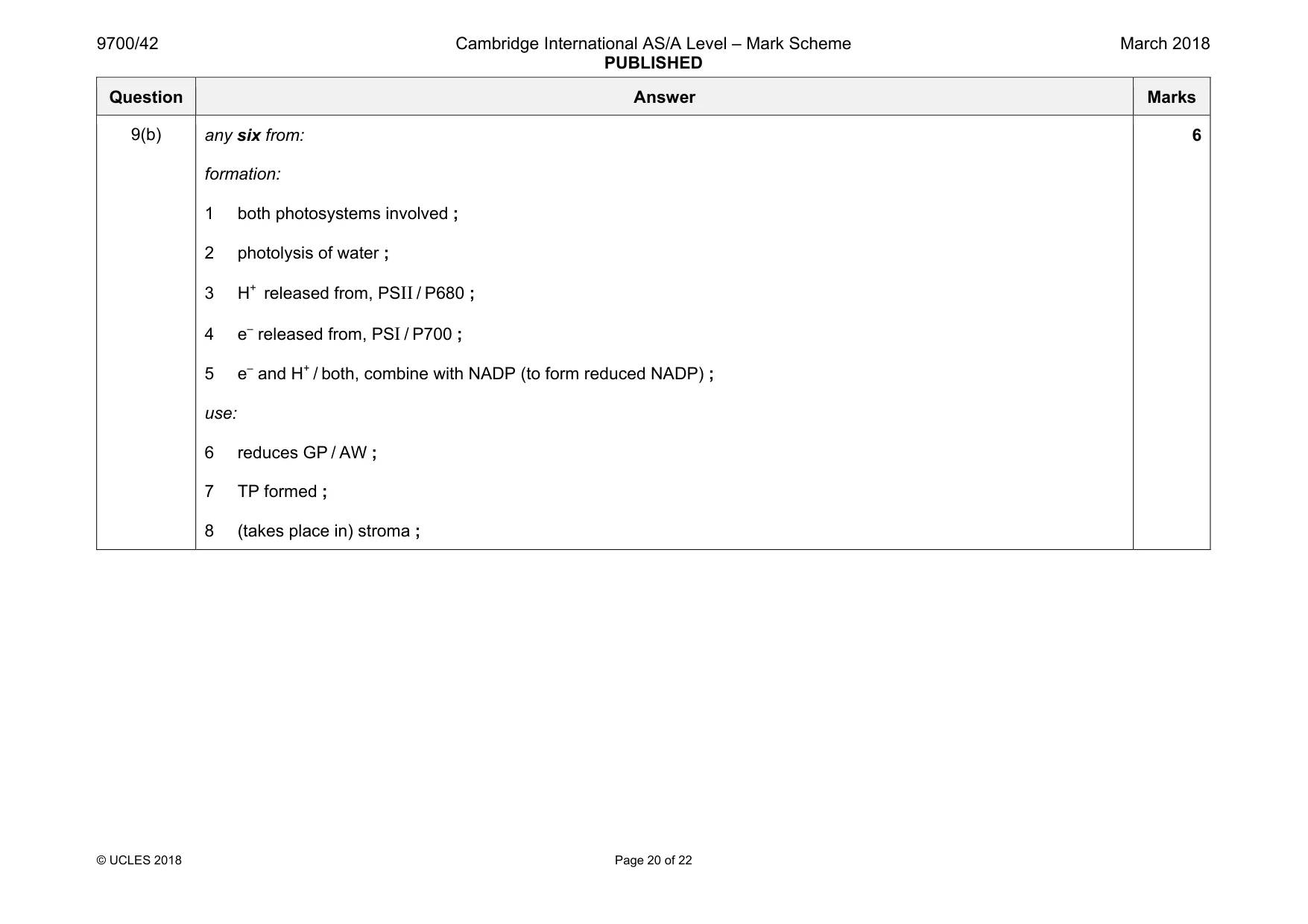
9 (a) Describe the process of cyclic photophosphorylation and the structure of the photosystem involved. [9] (b) Explain how non-cyclic photophosphorylation produces reduced NADP and how reduced NADP is used in the light independent stage. [6] [Total: 15]
Mark scheme: 9(a) any nine from: cyclic photophosphorylation: 1 (only) PSI / P700, involved ; 2 light energy absorbed ; 3 (results in) electron excited / AW ; 4 (electron) emitted from chlorophyll ; 5 chain of electron carriers / ETC ; 6 ATP synthesis ; 7 electron returns to, PSI / P700 ; photosystems: 8 pigments arranged in light-harvesting clusters ; 9 primary pigment / chlorophyll a / reaction centre ; 10 accessory pigments / chlorophyll b / carotenoids, surround, primary pigment / reaction centre / chlorophyll a ; 11 photosystem located in thylakoid ; 9 Question Answer Marks 9(b) any six from: formation: 1 both photosystems involved ; 2 photolysis of water ; 3 H+ released from, PSII / P680 ; 4 e– released from, PSI / P700 ; 5 e– and H+ / both, combine with NADP (to form reduced NADP) ; use: 6 reduces GP / AW ; 7 TP formed ; 8 (takes place in) stroma ; 6
More questions on Photosynthesis as an energy transfer process
Q10 · Explain the role of auxin in cell elongation
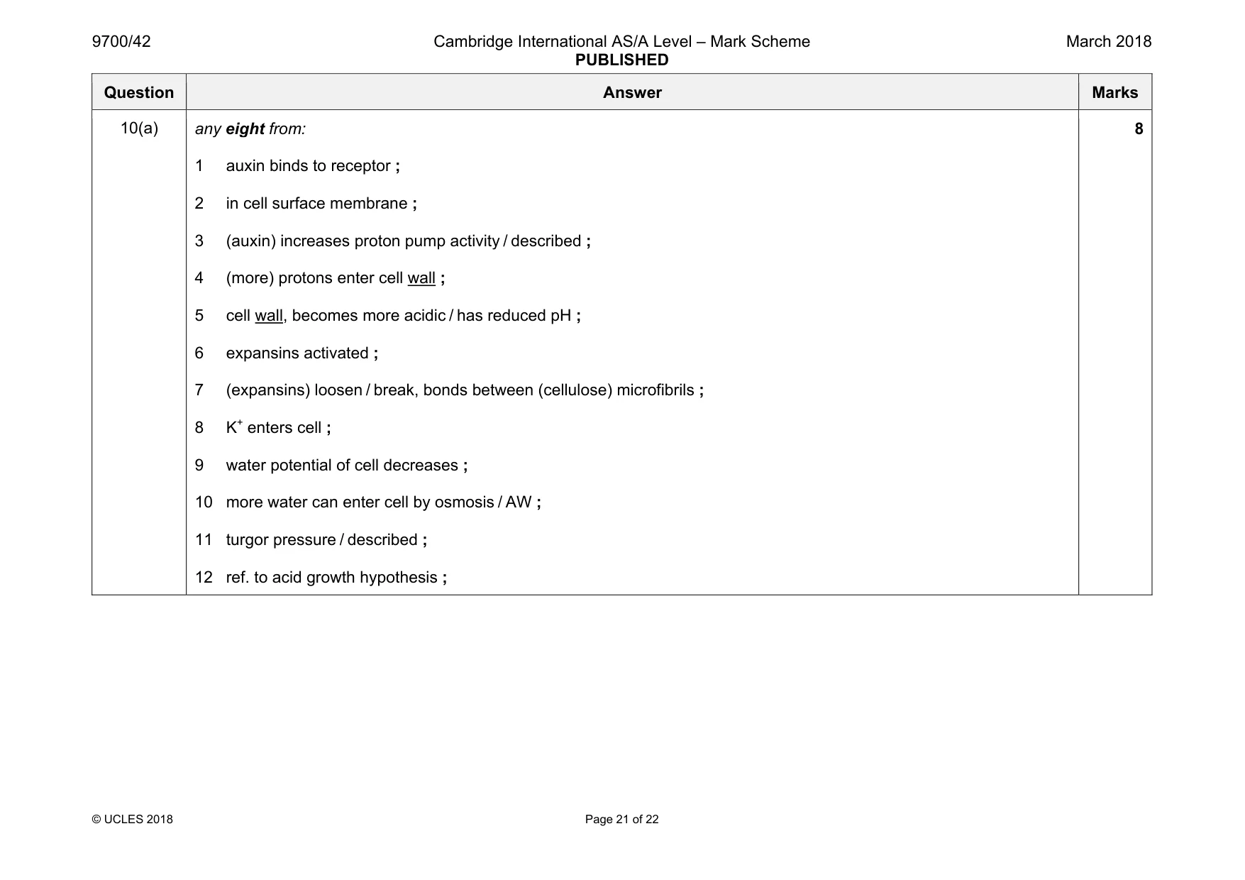
10 (a) Explain the role of auxin in cell elongation. [8] (b) Explain, using examples, how the environment may affect the phenotype of individual organisms. [7] [Total: 15] .................................................................................................................................................................. .................................................................................................................................................................. .................................................................................................................................................................. .................................................................................................................................................................. .................................................................................................................................................................. .................................................................................................................................................................. .................................................................................................................................................................. .................................................................................................................................................................. .................................................................................................................................................................. .................................................................................................................................................................. .................................................................................................................................................................. .................................................................................................................................................................. .................................................................................................................................................................. .................................................................................................................................................................. .................................................................................................................................................................. .................................................................................................................................................................. .................................................................................................................................................................. .................................................................................................................................................................. .................................................................................................................................................................. .................................................................................................................................................................. .................................................................................................................................................................. .................................................................................................................................................................. .................................................................................................................................................................. .................................................................................................................................................................. .................................................................................................................................................................. .................................................................................................................................................................. .................................................................................................................................................................. .................................................................................................................................................................. .................................................................................................................................................................. .................................................................................................................................................................. .................................................................................................................................................................. .................................................................................................................................................................. .................................................................................................................................................................. .................................................................................................................................................................. .................................................................................................................................................................. .................................................................................................................................................................. .................................................................................................................................................................. .................................................................................................................................................................. .................................................................................................................................................................. .................................................................................................................................................................. .................................................................................................................................................................. .................................................................................................................................................................. .................................................................................................................................................................. .................................................................................................................................................................. .................................................................................................................................................................. .................................................................................................................................................................. .................................................................................................................................................................. .................................................................................................................................................................. .................................................................................................................................................................. .................................................................................................................................................................. .................................................................................................................................................................. .................................................................................................................................................................. .................................................................................................................................................................. .................................................................................................................................................................. .................................................................................................................................................................. .................................................................................................................................................................. .................................................................................................................................................................. .................................................................................................................................................................. .................................................................................................................................................................. .................................................................................................................................................................. .................................................................................................................................................................. .................................................................................................................................................................. .................................................................................................................................................................. .................................................................................................................................................................. .................................................................................................................................................................. .................................................................................................................................................................. .................................................................................................................................................................. .................................................................................................................................................................. .................................................................................................................................................................. .................................................................................................................................................................. .................................................................................................................................................................. .................................................................................................................................................................. ..................................................................................................................................................................
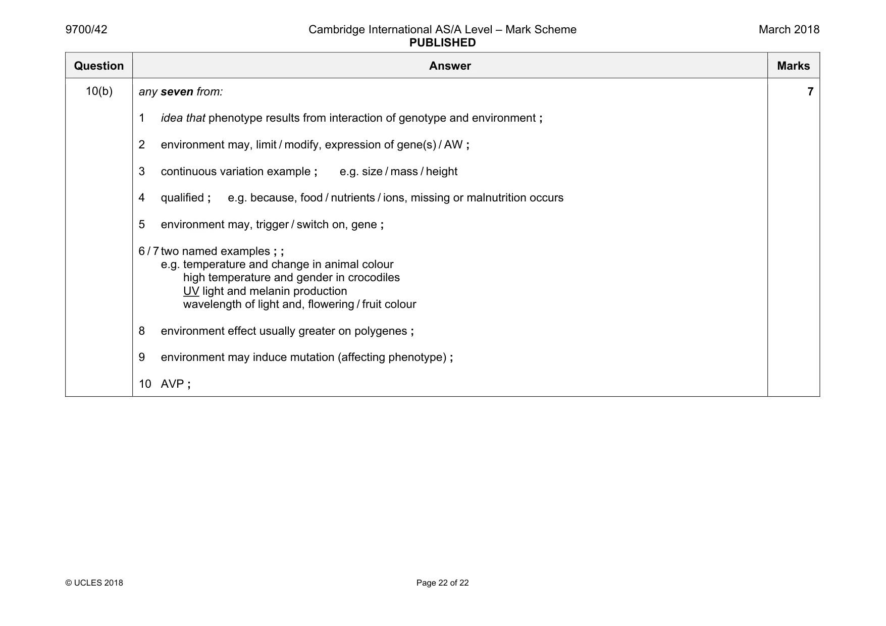
Mark scheme: 10(a) any eight from: 1 auxin binds to receptor ; 2 in cell surface membrane ; 3 (auxin) increases proton pump activity / described ; 4 (more) protons enter cell wall ; 5 cell wall, becomes more acidic / has reduced pH ; 6 expansins activated ; 7 (expansins) loosen / break, bonds between (cellulose) microfibrils ; 8 K+ enters cell ; 9 water potential of cell decreases ; 10 more water can enter cell by osmosis / AW ; 11 turgor pressure / described ; 12 ref. to acid growth hypothesis ; 8 Question Answer Marks 10(b) any seven from: 1 idea that phenotype results from interaction of genotype and environment ; 2 environment may, limit / modify, expression of gene(s) / AW ; 3 continuous variation example ; e.g. size / mass / height 4 qualified ; e.g. because, food / nutrients / ions, missing or malnutrition occurs 5 environment may, trigger / switch on, gene ; 6 / 7 two named examples ; ; e.g. temperature and change in animal colour high temperature and gender in crocodiles UV light and melanin production wavelength of light and, flowering / fruit colour 8 environment effect usually greater on polygenes ; 9 environment may induce mutation (affecting phenotype) ; 10 AVP ; 7
What was in this paper
The subtopics covered by these 10 questions, and how many questions each got. Open one in a new tab to see every Cambridge question on it.
What you needed in this session
Cambridge’s own grade thresholds for 2018 Feb/March, Paper 4 · Variant 2. A higher threshold means an easier paper — the bar moves with how the cohort did.