Cambridge A Level Biology 9700 — 2016 May/June Paper 3 · Variant 1
9700/31/M/J/16 · 40 marks · ≈45 min
The question paper and its mark scheme, free to read here and free to download. This is Cambridge’s own paper, exactly as it was sat.
Question paper12 pages

Mark scheme
No mark scheme is in the library for this paper.
Paper as text
Question paper, page 1
This document consists of 11 printed pages and 1 blank page. DC (ST/SW) 105810/3 © UCLES 2016 [Turn over Cambridge International Examinations Cambridge International Advanced Subsidiary and Advanced Level * 5 4 0 6 4 9 7 8 3 4 * BIOLOGY 9700/31 Paper 3 Advanced Practical Skills 1 May/June 2016 2 hours Candidates answer on the Question Paper. Additional Materials: As listed in the Confidential Instructions. READ THESE INSTRUCTIONS FIRST Write your Centre number, candidate number and name on all the work you hand in. Write in dark blue or black pen. You may use an HB pencil for any diagrams or graphs. Do not use staples, paper clips, glue or correction fluid. DO NOT WRITE IN ANY BARCODES. Answer all questions. Electronic calculators may be used. You may lose marks if you do not show your working or if you do not use appropriate units. At the end of the examination, fasten all your work securely together. The number of marks is given in brackets [ ] at the end of each question or part question. For Examiner’s Use 1 2 Total
Question paper, page 2
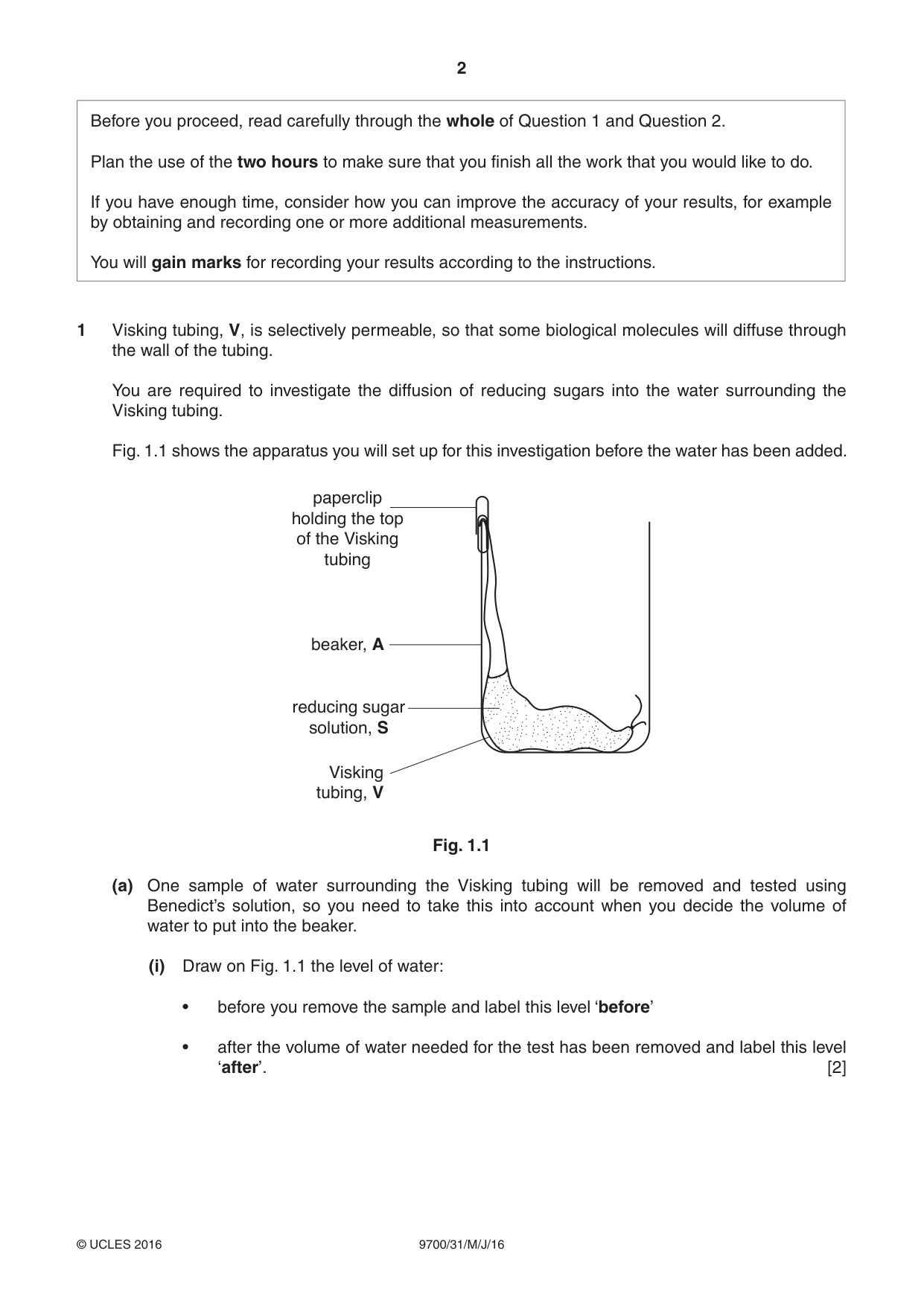
2 9700/31/M/J/16 © UCLES 2016 Before you proceed, read carefully through the whole of Question 1 and Question 2. Plan the use of the two hours to make sure that you finish all the work that you would like to do. If you have enough time, consider how you can improve the accuracy of your results, for example by obtaining and recording one or more additional measurements. You will gain marks for recording your results according to the instructions. 1 Visking tubing, V, is selectively permeable, so that some biological molecules will diffuse through the wall of the tubing. You are required to investigate the diffusion of reducing sugars into the water surrounding the Visking tubing. Fig. 1.1 shows the apparatus you will set up for this investigation before the water has been added. paperclip holding the top of the Visking tubing beaker, A reducing sugar solution, S Visking tubing, V Fig. 1.1 (a) One sample of water surrounding the Visking tubing will be removed and tested using Benedict’s solution, so you need to take this into account when you decide the volume of water to put into the beaker. (i) Draw on Fig. 1.1 the level of water: • before you remove the sample and label this level ‘before’ • after the volume of water needed for the test has been removed and label this level ‘after’. [2]
Question paper, page 3
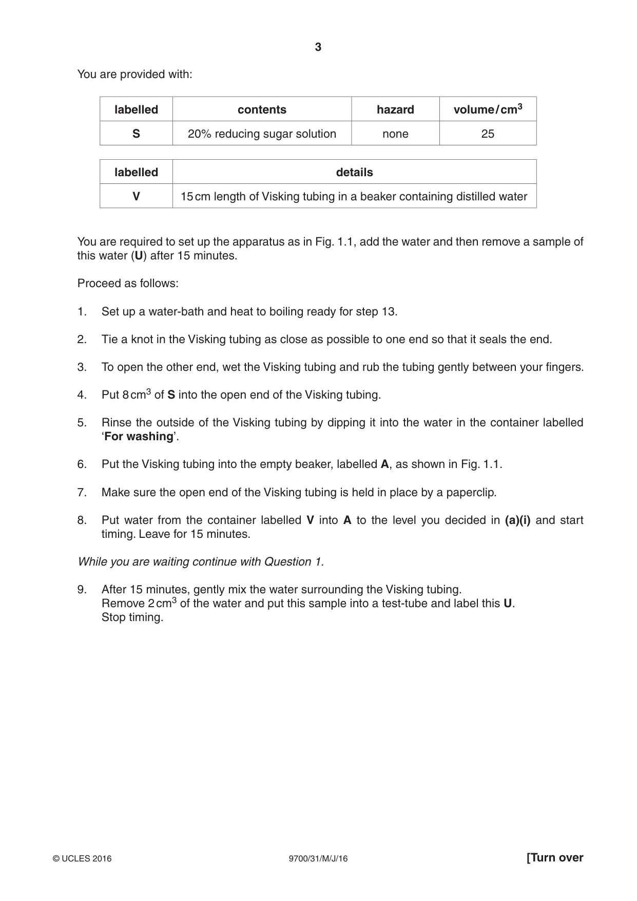
3 9700/31/M/J/16 © UCLES 2016 [Turn over You are provided with: labelled contents hazard volume / cm3 S 20% reducing sugar solution none 25 labelled details V 15 cm length of Visking tubing in a beaker containing distilled water You are required to set up the apparatus as in Fig. 1.1, add the water and then remove a sample of this water (U) after 15 minutes. Proceed as follows: 1. Set up a water-bath and heat to boiling ready for step 13. 2. Tie a knot in the Visking tubing as close as possible to one end so that it seals the end. 3. To open the other end, wet the Visking tubing and rub the tubing gently between your fingers. 4. Put 8 cm3 of S into the open end of the Visking tubing. 5. Rinse the outside of the Visking tubing by dipping it into the water in the container labelled ‘For washing’. 6. Put the Visking tubing into the empty beaker, labelled A, as shown in Fig. 1.1. 7. Make sure the open end of the Visking tubing is held in place by a paperclip. 8. Put water from the container labelled V into A to the level you decided in (a)(i) and start timing. Leave for 15 minutes. While you are waiting continue with Question 1. 9. After 15 minutes, gently mix the water surrounding the Visking tubing. Remove 2 cm3 of the water and put this sample into a test-tube and label this U. Stop timing.
Question paper, page 4
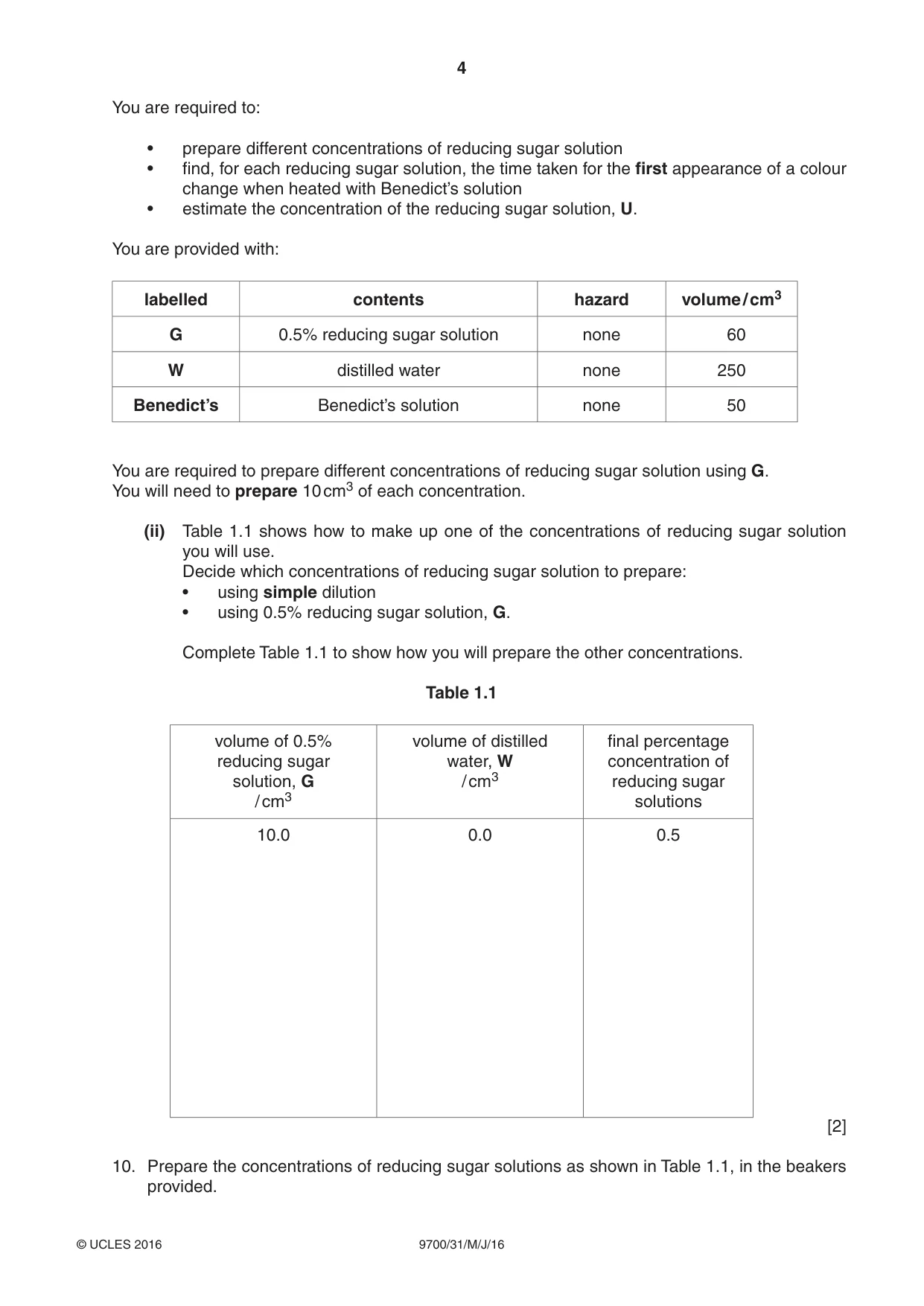
4 9700/31/M/J/16 © UCLES 2016 You are required to: • prepare different concentrations of reducing sugar solution • find, for each reducing sugar solution, the time taken for the first appearance of a colour change when heated with Benedict’s solution • estimate the concentration of the reducing sugar solution, U. You are provided with: labelled contents hazard volume / cm3 G 0.5% reducing sugar solution none 60 W distilled water none 250 Benedict’s Benedict’s solution none 50 You are required to prepare different concentrations of reducing sugar solution using G. You will need to prepare 10 cm3 of each concentration. (ii) Table 1.1 shows how to make up one of the concentrations of reducing sugar solution you will use. Decide which concentrations of reducing sugar solution to prepare: • using simple dilution • using 0.5% reducing sugar solution, G. Complete Table 1.1 to show how you will prepare the other concentrations. Table 1.1 volume of 0.5% reducing sugar solution, G / cm3 volume of distilled water, W / cm3 final percentage concentration of reducing sugar solutions 10.0 0.0 0.5 [2] 10. Prepare the concentrations of reducing sugar solutions as shown in Table 1.1, in the beakers provided.
Question paper, page 5
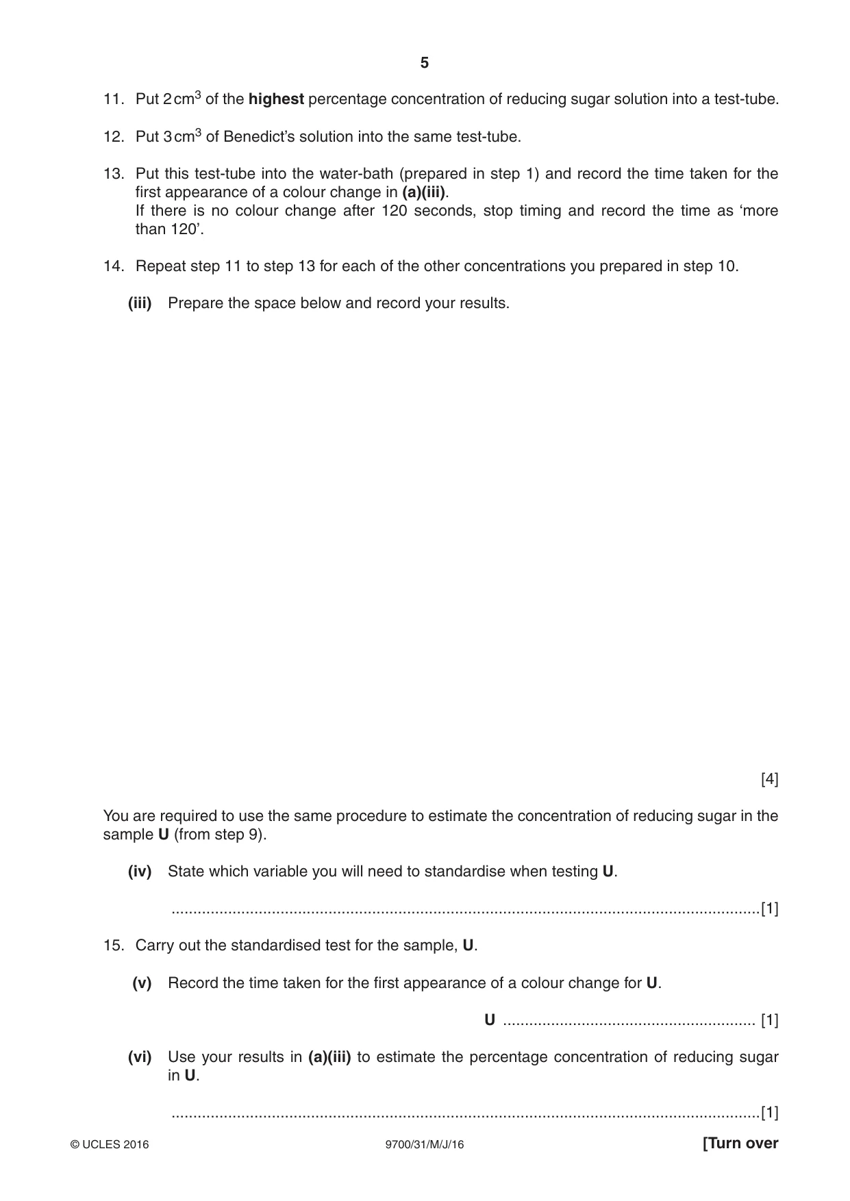
5 9700/31/M/J/16 © UCLES 2016 [Turn over 11. Put 2 cm3 of the highest percentage concentration of reducing sugar solution into a test-tube. 12. Put 3 cm3 of Benedict’s solution into the same test-tube. 13. Put this test-tube into the water-bath (prepared in step 1) and record the time taken for the first appearance of a colour change in (a)(iii). If there is no colour change after 120 seconds, stop timing and record the time as ‘more than 120’. 14. Repeat step 11 to step 13 for each of the other concentrations you prepared in step 10. (iii) Prepare the space below and record your results. [4] You are required to use the same procedure to estimate the concentration of reducing sugar in the sample U (from step 9). (iv) State which variable you will need to standardise when testing U. …[1] 15. Carry out the standardised test for the sample, U. (v) Record the time taken for the first appearance of a colour change for U. U … [1] (vi) Use your results in (a)(iii) to estimate the percentage concentration of reducing sugar in U. …[1]
Question paper, page 6
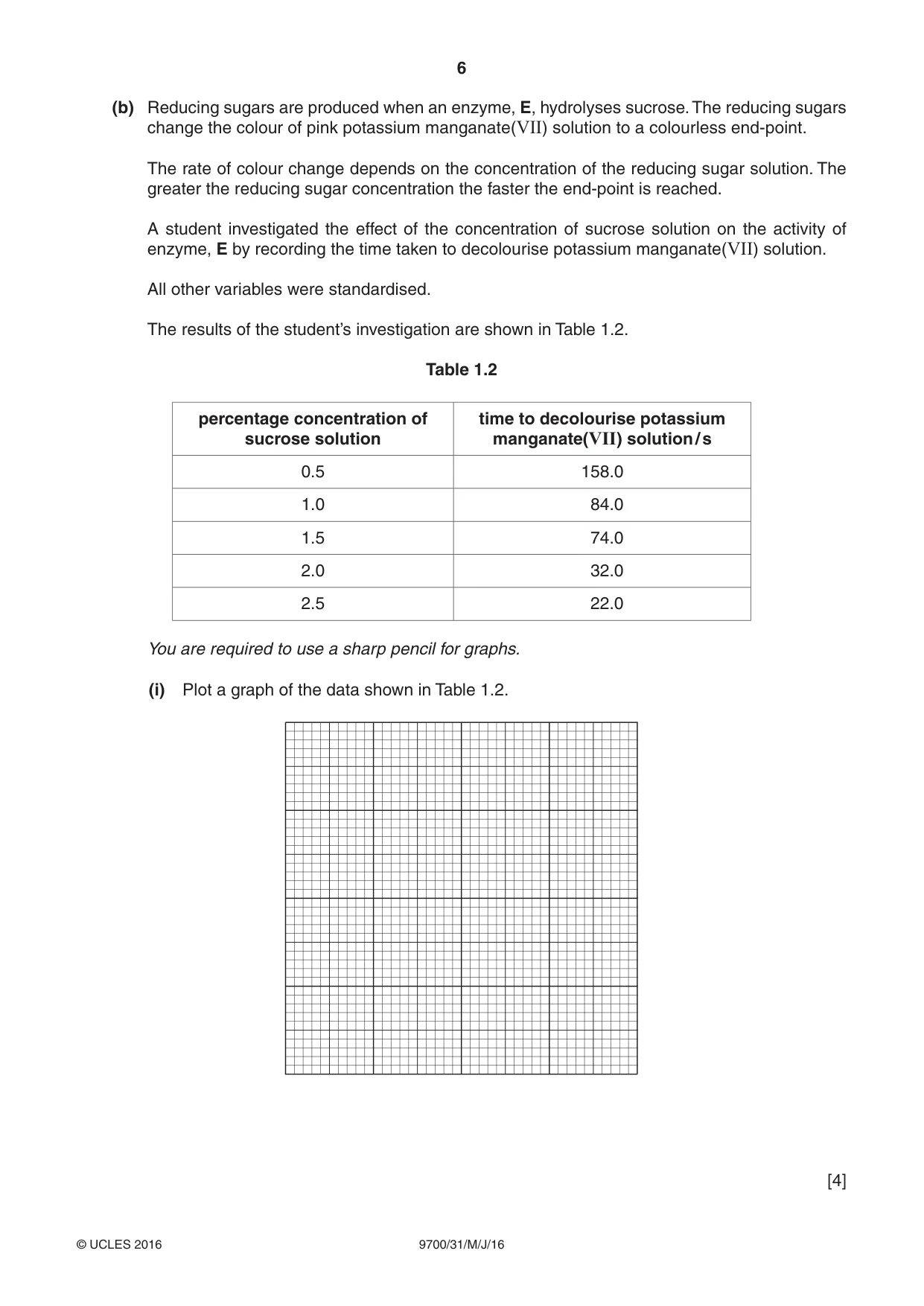
6 9700/31/M/J/16 © UCLES 2016 (b) Reducing sugars are produced when an enzyme, E, hydrolyses sucrose. The reducing sugars change the colour of pink potassium manganate(VII) solution to a colourless end-point. The rate of colour change depends on the concentration of the reducing sugar solution. The greater the reducing sugar concentration the faster the end-point is reached. A student investigated the effect of the concentration of sucrose solution on the activity of enzyme, E by recording the time taken to decolourise potassium manganate(VII) solution. All other variables were standardised. The results of the student’s investigation are shown in Table 1.2. Table 1.2 percentage concentration of sucrose solution time to decolourise potassium manganate(VII) solution / s 0.5 158.0 1.0 84.0 1.5 74.0 2.0 32.0 2.5 22.0 You are required to use a sharp pencil for graphs. (i) Plot a graph of the data shown in Table 1.2. [4]
Question paper, page 7
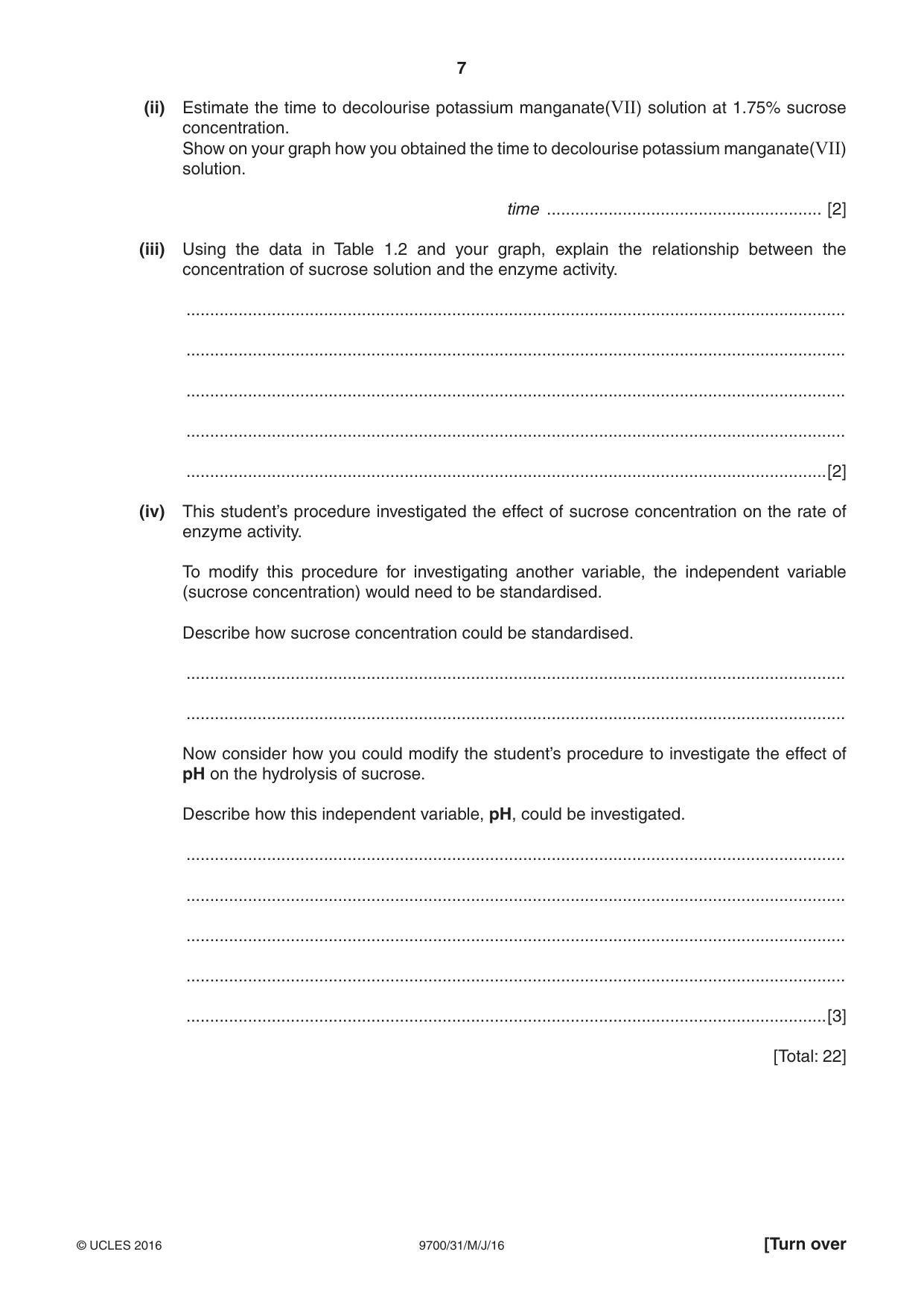
7 9700/31/M/J/16 © UCLES 2016 [Turn over (ii) Estimate the time to decolourise potassium manganate(VII) solution at 1.75% sucrose concentration. Show on your graph how you obtained the time to decolourise potassium manganate(VII) solution. time … [2] (iii) Using the data in Table 1.2 and your graph, explain the relationship between the concentration of sucrose solution and the enzyme activity. … … … … …[2] (iv) This student’s procedure investigated the effect of sucrose concentration on the rate of enzyme activity. To modify this procedure for investigating another variable, the independent variable (sucrose concentration) would need to be standardised. Describe how sucrose concentration could be standardised. … … Now consider how you could modify the student’s procedure to investigate the effect of pH on the hydrolysis of sucrose. Describe how this independent variable, pH, could be investigated. … … … … …[3] [Total: 22]
Question paper, page 8
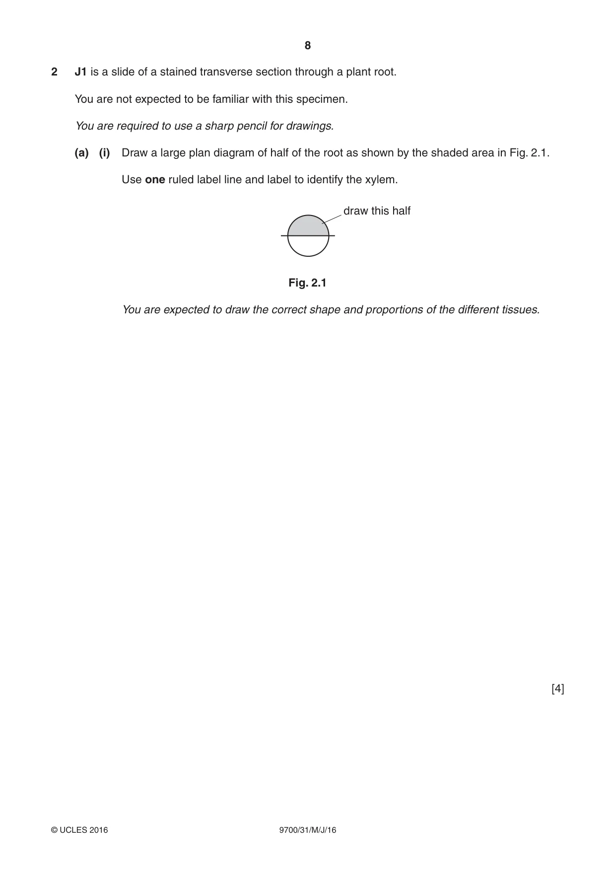
8 9700/31/M/J/16 © UCLES 2016 2 J1 is a slide of a stained transverse section through a plant root. You are not expected to be familiar with this specimen. You are required to use a sharp pencil for drawings. (a) (i) Draw a large plan diagram of half of the root as shown by the shaded area in Fig. 2.1. Use one ruled label line and label to identify the xylem. draw this half Fig. 2.1 You are expected to draw the correct shape and proportions of the different tissues. [4]
Question paper, page 9
9 9700/31/M/J/16 © UCLES 2016 [Turn over (ii) Observe the vascular tissue in the centre of the root on J1. The vascular tissue is surrounded by a ring of cells. Select one group of four adjacent (touching) cells from this ring of cells. Each cell of the group must touch at least one of the other cells. Make a large drawing of this group of four cells. Use one ruled label line and label to identify the cell wall of one cell. [5]
Question paper, page 10
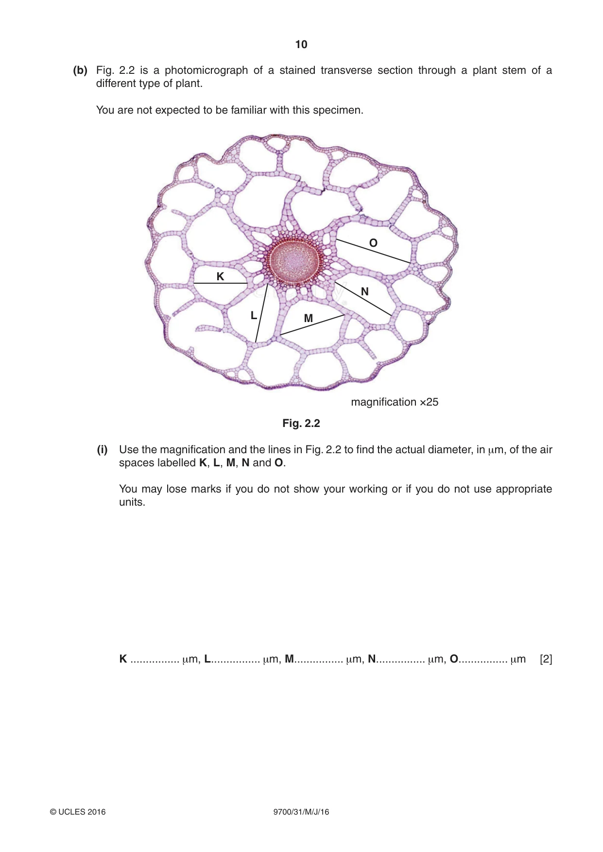
10 9700/31/M/J/16 © UCLES 2016 (b) Fig. 2.2 is a photomicrograph of a stained transverse section through a plant stem of a different type of plant. You are not expected to be familiar with this specimen. K L M N O magnification ×25 Fig. 2.2 (i) Use the magnification and the lines in Fig. 2.2 to find the actual diameter, in μm, of the air spaces labelled K, L, M, N and O. You may lose marks if you do not show your working or if you do not use appropriate units. K … μm, L… μm, M… μm, N… μm, O… μm [2]
Question paper, page 11
11 9700/31/M/J/16 © UCLES 2016 (ii) Using the actual diameters calculated in (b)(i), calculate the mean actual diameter of an air space. You may lose marks if you do not show your working or if you do not use appropriate units. mean actual diameter … [2] (iii) A feature of the plant in Fig. 2.2 is the presence of air spaces in the plant stem. Suggest a habitat where this plant might grow … Suggest how the air spaces adapt the plant to this habitat. … …[1] (c) Prepare the space below so that it is suitable for you to record the observable differences between the root on J1 and the stem in Fig. 2.2. Record your observations in the space you have prepared. [4] [Total: 18]
Question paper, page 12
12 9700/31/M/J/16 © UCLES 2016 Permission to reproduce items where third-party owned material protected by copyright is included has been sought and cleared where possible. Every reasonable effort has been made by the publisher (UCLES) to trace copyright holders, but if any items requiring clearance have unwittingly been included, the publisher will be pleased to make amends at the earliest possible opportunity. To avoid the issue of disclosure of answer-related information to candidates, all copyright acknowledgements are reproduced online in the Cambridge International Examinations Copyright Acknowledgements Booklet. This is produced for each series of examinations and is freely available to download at www.cie.org.uk after the live examination series. Cambridge International Examinations is part of the Cambridge Assessment Group. Cambridge Assessment is the brand name of University of Cambridge Local Examinations Syndicate (UCLES), which is itself a department of the University of Cambridge. BLANK PAGE
What you needed in this session
Cambridge’s own grade thresholds for 2016 May/June, Paper 3 · Variant 1. A higher threshold means an easier paper — the bar moves with how the cohort did.