Cambridge A Level Biology 9700 — 2005 Oct/Nov Paper 5 · Variant 1
9700/51/O/N/05
The question paper and its mark scheme, free to read here and free to download. This is Cambridge’s own paper, exactly as it was sat.
Question paper8 pages

Mark scheme3 pages
Answers below. Sit the paper first if you are practising.
Paper as text
Question paper, page 1
This document consists of 7 printed pages and 1 blank page. SP (CW) S85327/2 © UCLES 2005 [Turn over UNIVERSITY OF CAMBRIDGE INTERNATIONAL EXAMINATIONS General Certificate of Education Advanced Level BIOLOGY 9700/05 Paper 5 Practical Test A2 October/November 2005 1 hour 30 minutes Candidates answer on the Question Paper. Additional Materials: As listed in Instructions to Supervisors. READ THESE INSTRUCTIONS FIRST Write your Centre number, candidate number and name in the spaces provided at the top of this page. Write in dark blue or black pen in the spaces provided on the Question Paper. You may use a soft pencil for any diagrams, graphs or rough working. Do not use staples, paper clips, highlighters, glue or correction fluid. Answer all questions. The number of marks is given in brackets [ ] at the end of each question or part question. You are advised to spend 50 minutes on Question 1 and 40 minutes on Question 2. Centre Number Candidate Number Name For Examiner’s Use 1 2 Total
Question paper, page 2
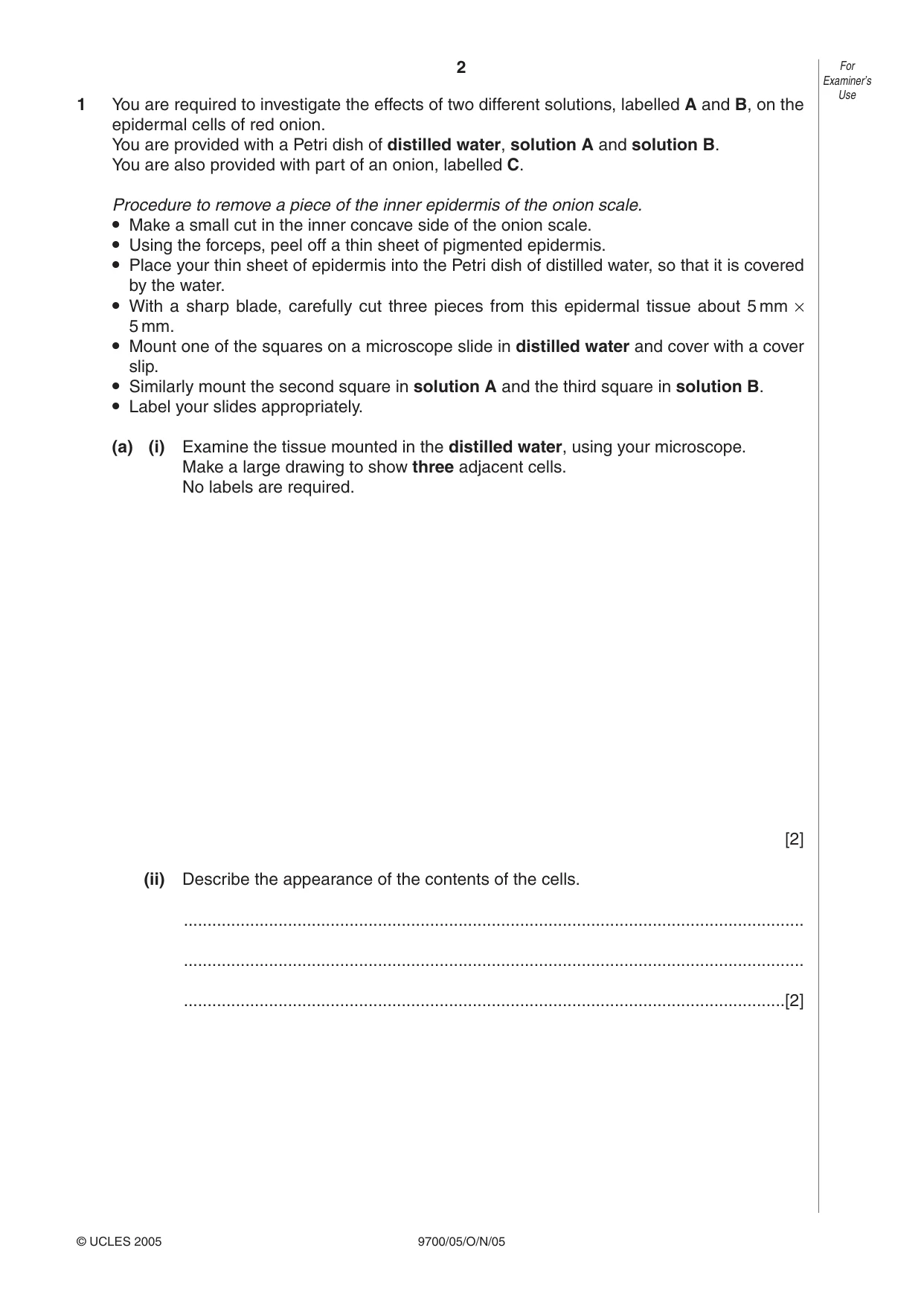
2 9700/05/O/N/05 1 You are required to investigate the effects of two different solutions, labelled A and B, on the epidermal cells of red onion. You are provided with a Petri dish of distilled water, solution A and solution B. You are also provided with part of an onion, labelled C. Procedure to remove a piece of the inner epidermis of the onion scale. • Make a small cut in the inner concave side of the onion scale. • Using the forceps, peel off a thin sheet of pigmented epidermis. • Place your thin sheet of epidermis into the Petri dish of distilled water, so that it is covered by the water. • With a sharp blade, carefully cut three pieces from this epidermal tissue about 5 mm × 5 mm. • Mount one of the squares on a microscope slide in distilled water and cover with a cover slip. • Similarly mount the second square in solution A and the third square in solution B. • Label your slides appropriately. (a) (i) Examine the tissue mounted in the distilled water, using your microscope. Make a large drawing to show three adjacent cells. No labels are required. [2] (ii) Describe the appearance of the contents of the cells. … … …[2] For Examiner’s Use © UCLES 2005
Question paper, page 3
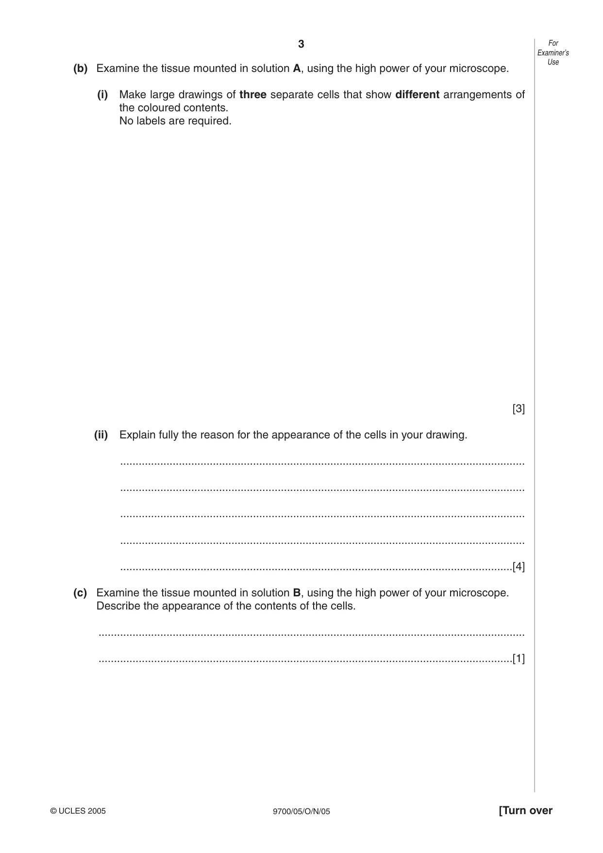
3 9700/05/O/N/05 (b) Examine the tissue mounted in solution A, using the high power of your microscope. (i) Make large drawings of three separate cells that show different arrangements of the coloured contents. No labels are required. [3] (ii) Explain fully the reason for the appearance of the cells in your drawing. … … … … …[4] (c) Examine the tissue mounted in solution B, using the high power of your microscope. Describe the appearance of the contents of the cells. … …[1] For Examiner’s Use © UCLES 2005 [Turn over
Question paper, page 4
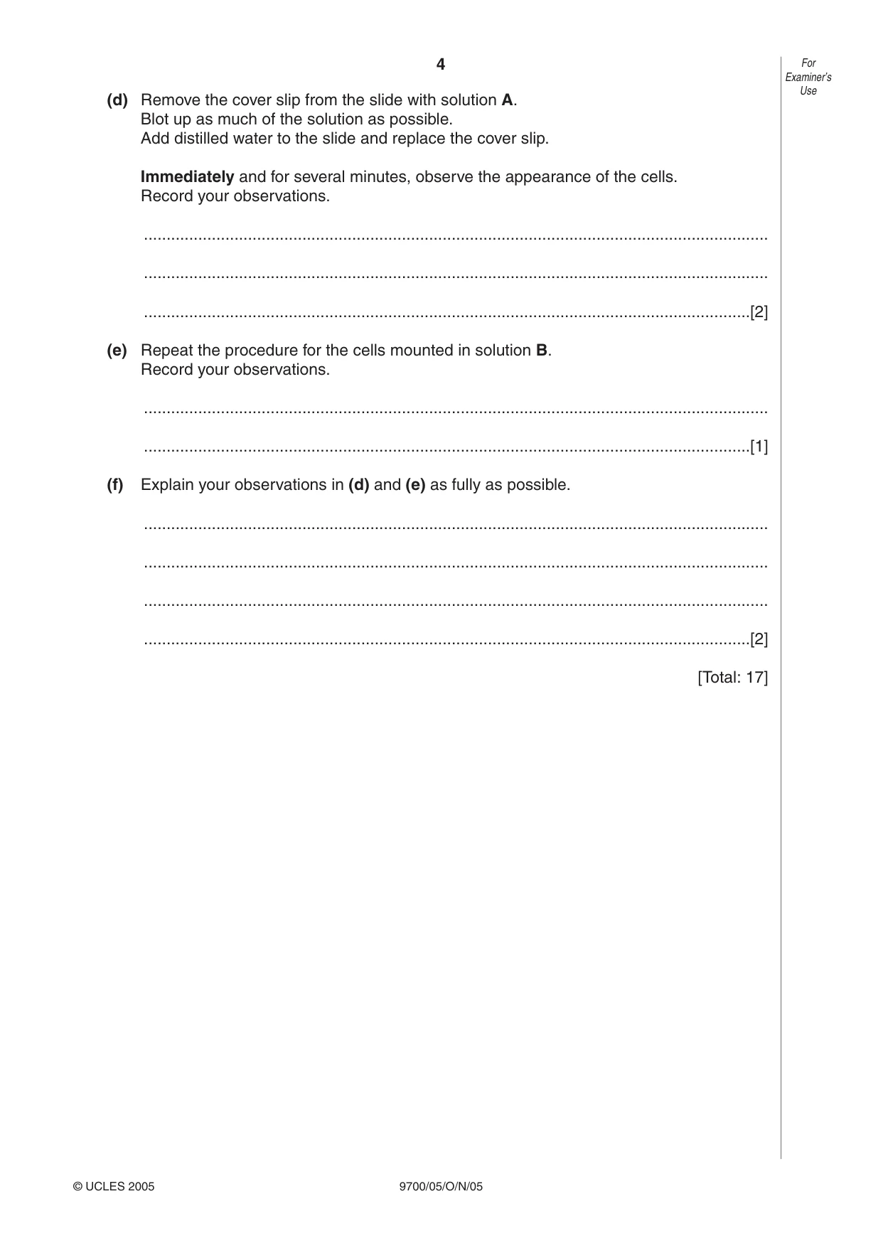
4 9700/05/O/N/05 (d) Remove the cover slip from the slide with solution A. Blot up as much of the solution as possible. Add distilled water to the slide and replace the cover slip. Immediately and for several minutes, observe the appearance of the cells. Record your observations. … … …[2] (e) Repeat the procedure for the cells mounted in solution B. Record your observations. … …[1] (f) Explain your observations in (d) and (e) as fully as possible. … … … …[2] [Total: 17] For Examiner’s Use © UCLES 2005
Question paper, page 5
5 9700/05/O/N/05 BLANK PAGE Question 2 is on page 6. [Turn over
Question paper, page 6
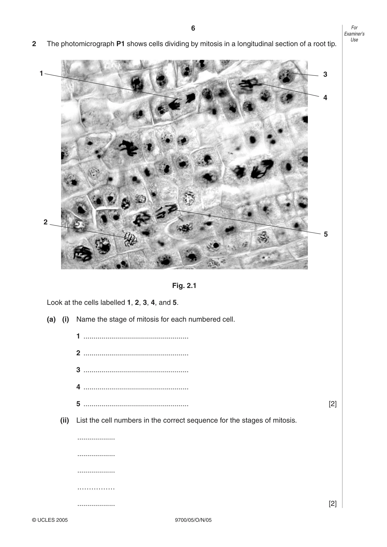
6 9700/05/O/N/05 For Examiner’s Use © UCLES 2005 2 The photomicrograph P1 shows cells dividing by mitosis in a longitudinal section of a root tip. Fig. 2.1 Look at the cells labelled 1, 2, 3, 4, and 5. (a) (i) Name the stage of mitosis for each numbered cell. 1 … 2 … 3 … 4 … 5 … [2] (ii) List the cell numbers in the correct sequence for the stages of mitosis. … … … .…………… … [2] 1 2 5 3 4
Question paper, page 7
7 9700/05/O/N/05 (iii) Choose a cell at metaphase. Do not choose cells 1, 2, 3, 4 or 5. Make a large, labelled drawing of your chosen cell. Draw, on Fig. 2.1, a ring round your chosen cell. [4] Question 2 continues on page 8. For Examiner’s Use © UCLES 2005 [Turn over
Question paper, page 8
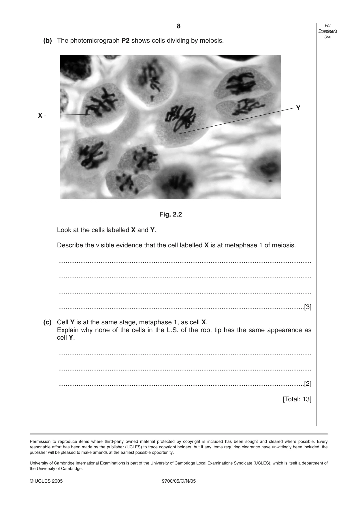
8 9700/05/O/N/05 (b) The photomicrograph P2 shows cells dividing by meiosis. Fig. 2.2 Look at the cells labelled X and Y. Describe the visible evidence that the cell labelled X is at metaphase 1 of meiosis. … … … …[3] (c) Cell Y is at the same stage, metaphase 1, as cell X. Explain why none of the cells in the L.S. of the root tip has the same appearance as cell Y. … … …[2] [Total: 13] For Examiner’s Use © UCLES 2005 X Y Permission to reproduce items where third-party owned material protected by copyright is included has been sought and cleared where possible. Every reasonable effort has been made by the publisher (UCLES) to trace copyright holders, but if any items requiring clearance have unwittingly been included, the publisher will be pleased to make amends at the earliest possible opportunity. University of Cambridge International Examinations is part of the University of Cambridge Local Examinations Syndicate (UCLES), which is itself a department of the University of Cambridge.
Mark scheme, page 1
UNIVERSITY OF CAMBRIDGE INTERNATIONAL EXAMINATIONS GCE A/AS Level MARK SCHEME for the November 2005 question paper 9700 BIOLOGY 9700/05 Paper 5 maximum raw mark 30 This mark scheme is published as an aid to teachers and students, to indicate the requirements of the examination. It shows the basis on which Examiners were initially instructed to award marks. It does not indicate the details of the discussions that took place at an Examiners’ meeting before marking began. Any substantial changes to the mark scheme that arose from these discussions will be recorded in the published Report on the Examination. All Examiners are instructed that alternative correct answers and unexpected approaches in candidates’ scripts must be given marks that fairly reflect the relevant knowledge and skills demonstrated. Mark schemes must be read in conjunction with the question papers and the Report on the Examination. The minimum marks in these components needed for various grades were previously published with these mark schemes, but are now instead included in the Report on the Examination for this session. • CIE will not enter into discussion or correspondence in connection with these mark schemes. CIE is publishing the mark schemes for the November 2005 question papers for most IGCSE and GCE Advanced Level and Advanced Subsidiary Level syllabuses and some Ordinary Level syllabuses
Mark scheme, page 2
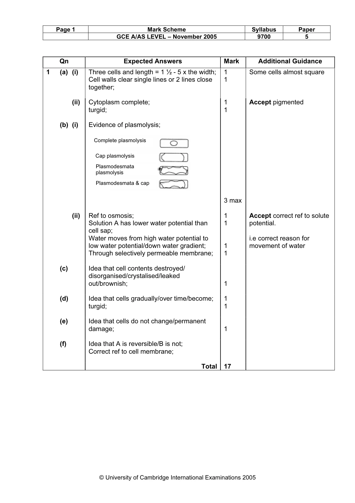
Page 1 Mark Scheme Syllabus Paper GCE A/AS LEVEL – November 2005 9700 5 © University of Cambridge International Examinations 2005 Qn Expected Answers Mark Additional Guidance 1 (a) (i) Three cells and length = 1 ½ - 5 x the width; Cell walls clear single lines or 2 lines close together; 1 1 Some cells almost square (ii) Cytoplasm complete; turgid; 1 1 Accept pigmented (b) (i) Evidence of plasmolysis; 3 max (ii) Ref to osmosis; Solution A has lower water potential than cell sap; Water moves from high water potential to low water potential/down water gradient; Through selectively permeable membrane; 1 1 1 1 Accept correct ref to solute potential. i.e correct reason for movement of water (c) Idea that cell contents destroyed/ disorganised/crystalised/leaked out/brownish; 1 (d) Idea that cells gradually/over time/become; turgid; 1 1 (e) Idea that cells do not change/permanent damage; 1 (f) Idea that A is reversible/B is not; Correct ref to cell membrane; Total 17 Complete plasmolysis Cap plasmolysis Plasmodesmata plasmolysis Plasmodesmata & cap
Mark scheme, page 3
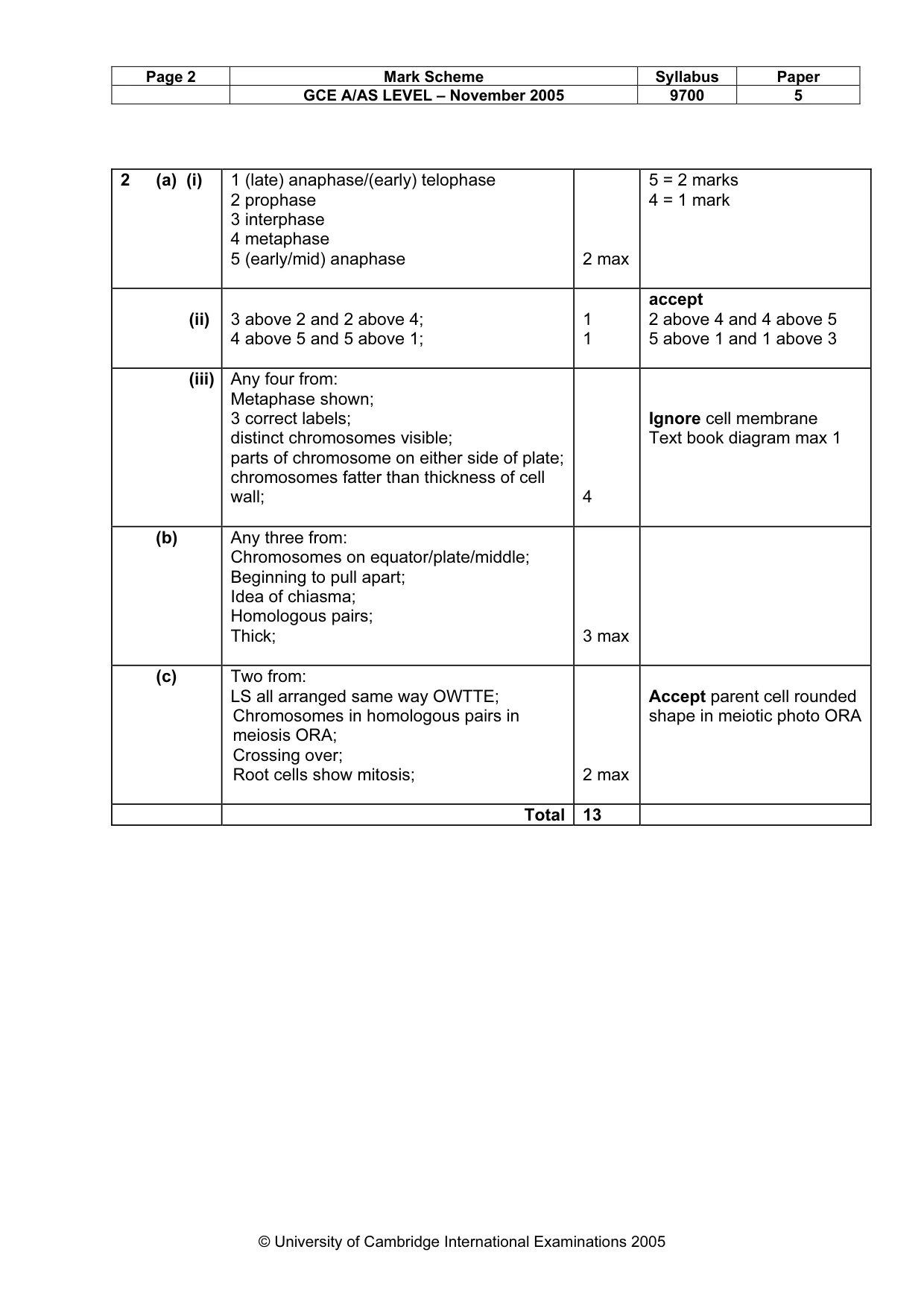
Page 2 Mark Scheme Syllabus Paper GCE A/AS LEVEL – November 2005 9700 5 © University of Cambridge International Examinations 2005 2 (a) (i) 1 (late) anaphase/(early) telophase 2 prophase 3 interphase 4 metaphase 5 (early/mid) anaphase 2 max 5 = 2 marks 4 = 1 mark (ii) 3 above 2 and 2 above 4; 4 above 5 and 5 above 1; 1 1 accept 2 above 4 and 4 above 5 5 above 1 and 1 above 3 (iii) Any four from: Metaphase shown; 3 correct labels; distinct chromosomes visible; parts of chromosome on either side of plate; chromosomes fatter than thickness of cell wall; 4 Ignore cell membrane Text book diagram max 1 (b) Any three from: Chromosomes on equator/plate/middle; Beginning to pull apart; Idea of chiasma; Homologous pairs; Thick; 3 max (c) Two from: LS all arranged same way OWTTE; Chromosomes in homologous pairs in meiosis ORA; Crossing over; Root cells show mitosis; 2 max Accept parent cell rounded shape in meiotic photo ORA Total 13