Cambridge A Level Biology 9700 — 2023 May/June Paper 3 · Variant 4
9700/34/M/J/23 · 2 questions · 40 marks · ≈45 min
The question paper and its mark scheme, free to read here and free to download. This is Cambridge’s own paper, exactly as it was sat.
Question paper16 pages
Mark scheme8 pages
Answers below. Sit the paper first if you are practising.
Questions as text
Q1 · Milk is a source of protein and reducing sugars
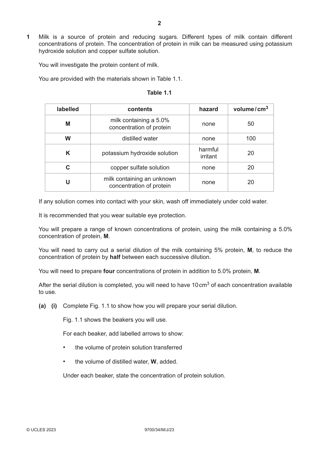
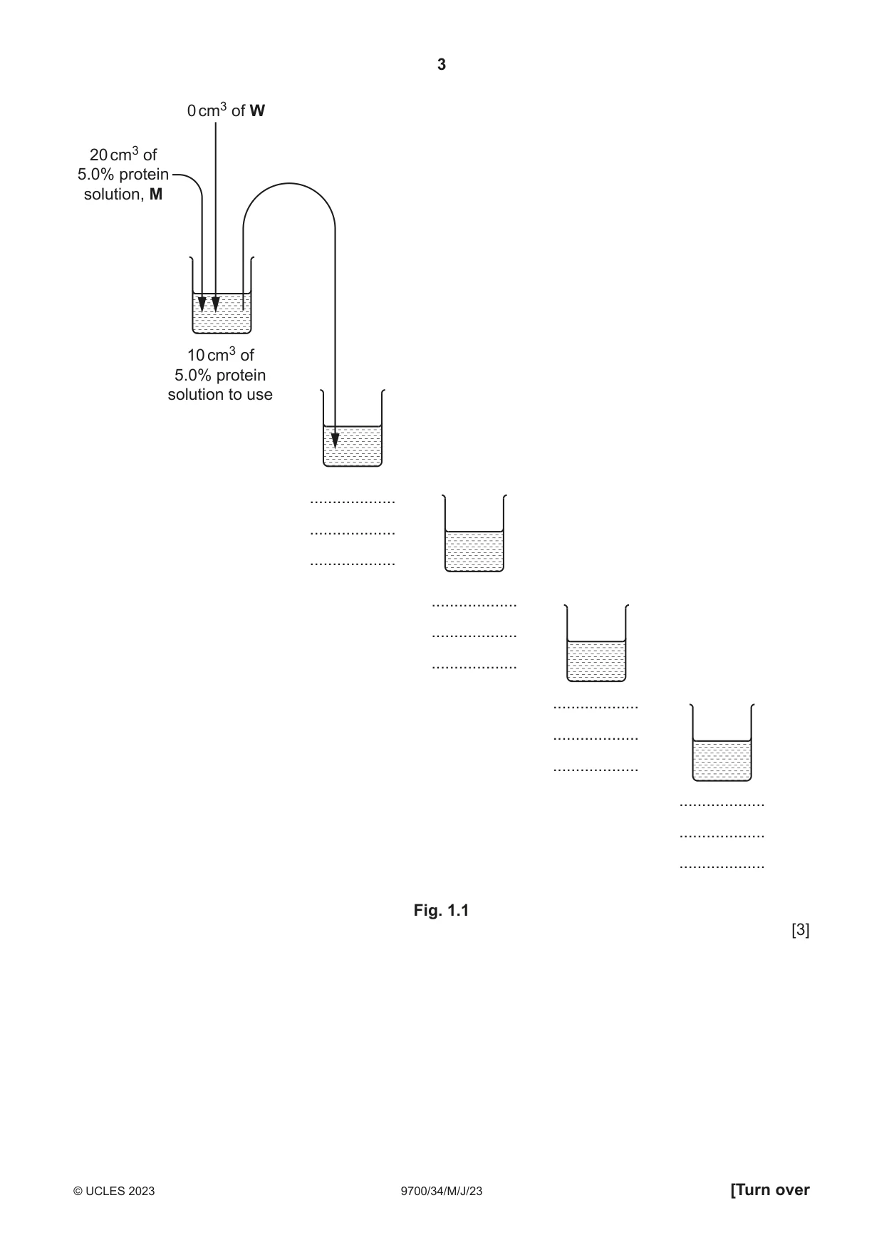
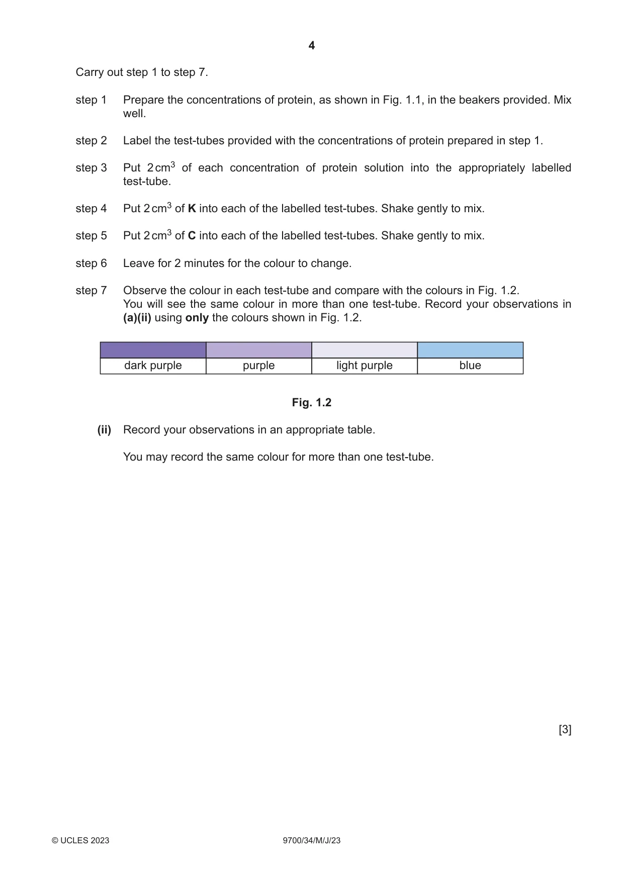
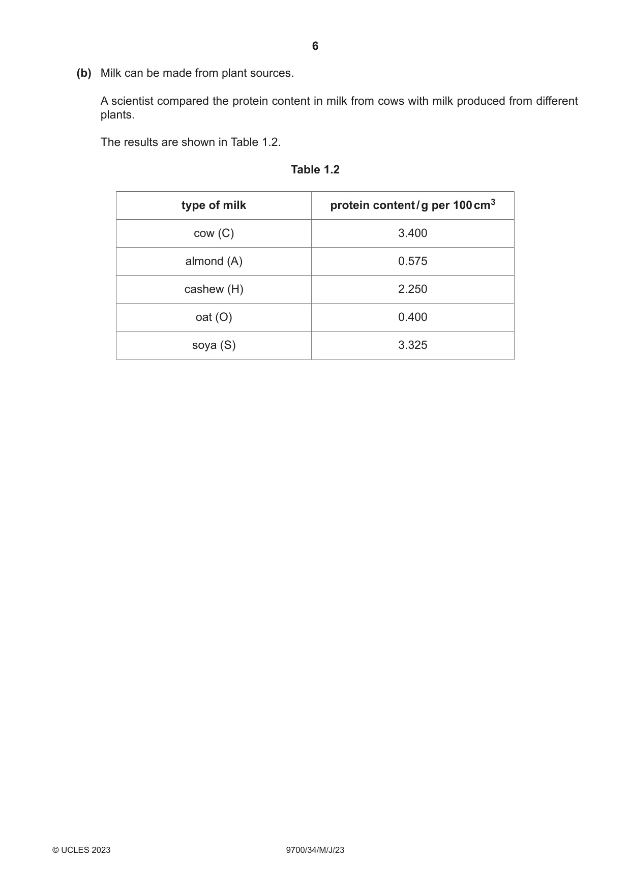
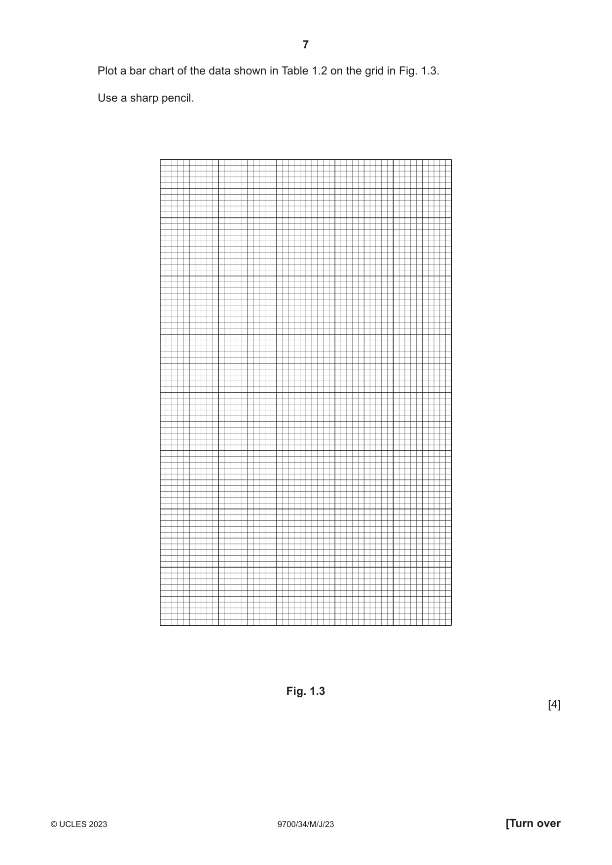
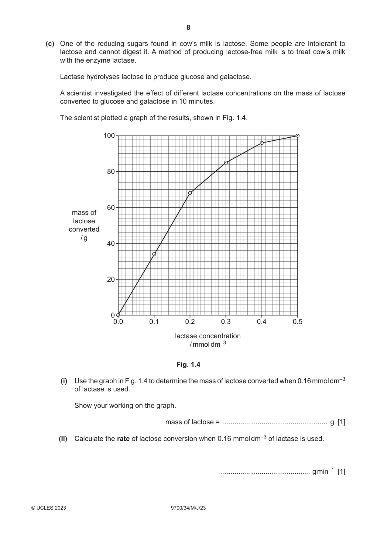
1 Milk is a source of protein and reducing sugars. Different types of milk contain different concentrations of protein. The concentration of protein in milk can be measured using potassium hydroxide solution and copper sulfate solution. You will investigate the protein content of milk. You are provided with the materials shown in Table 1.1. Table 1.1 labelled contents hazard volume / cm3 milk containing a 5.0% M none 50 concentration of protein W distilled water none 100 harmful K potassium hydroxide solution 20 irritant C copper sulfate solution none 20 milk containing an unknown U none 20 concentration of protein If any solution comes into contact with your skin, wash off immediately under cold water. It is recommended that you wear suitable eye protection. You will prepare a range of known concentrations of protein, using the milk containing a 5.0% concentration of protein, M. You will need to carry out a serial dilution of the milk containing 5% protein, M, to reduce the concentration of protein by half between each successive dilution. You will need to prepare four concentrations of protein in addition to 5.0% protein, M. After the serial dilution is completed, you will need to have 10 cm3 of each concentration available to use. (a) (i) Complete Fig. 1.1 to show how you will prepare your serial dilution. Fig. 1.1 shows the beakers you will use. For each beaker, add labelled arrows to show: • the volume of protein solution transferred • the volume of distilled water, W, added. Under each beaker, state the concentration of protein solution. 0 cm3 of W 20 cm3 of 5.0% protein solution, M 10 cm3 of 5.0% protein solution to use ................... ................... ................... ................... ................... ................... ................... ................... ................... ................... ................... ................... Fig. 1.1 [3] Carry out step 1 to step 7. step 1 Prepare the concentrations of protein, as shown in Fig. 1.1, in the beakers provided. Mix well. step 2 Label the test-tubes provided with the concentrations of protein prepared in step 1. step 3 Put 2 cm3 of each concentration of protein solution into the appropriately labelled test-tube. step 4 Put 2 cm3 of K into each of the labelled test-tubes. Shake gently to mix. step 5 Put 2 cm3 of C into each of the labelled test-tubes. Shake gently to mix. step 6 Leave for 2 minutes for the colour to change. step 7 Observe the colour in each test-tube and compare with the colours in Fig. 1.2. You will see the same colour in more than one test-tube. Record your observations in (a)(ii) using only the colours shown in Fig. 1.2. dark purple purple light purple blue Fig. 1.2 (ii) Record your observations in an appropriate table. You may record the same colour for more than one test-tube. [3] Carry out step 8 to step 9. step 8 Stir U and put 2 cm3 of U into a test-tube. step 9 Repeat step 4 to step 7 with U. Record the colour in (a)(iii). (iii) State the colour for sample U ...................................................................................... [1] (iv) Use your results in (a)(ii) and (a)(iii) to estimate the protein concentration in U. ..................................................................................................................................... [1] (v) Describe one significant source of error when carrying out step 7 and suggest an improvement to reduce this error. source of error ................................................................................................................... ........................................................................................................................................... improvement ..................................................................................................................... ........................................................................................................................................... [2] (vi) Suggest how you would modify the experiment to determine the concentration of reducing sugars in a sample of milk. ........................................................................................................................................... ........................................................................................................................................... ........................................................................................................................................... ........................................................................................................................................... ........................................................................................................................................... ........................................................................................................................................... ..................................................................................................................................... [3] (b) Milk can be made from plant sources. A scientist compared the protein content in milk from cows with milk produced from different plants. The results are shown in Table 1.2. Table 1.2 type of milk protein content / g per 100 cm3 cow (C) 3.400 almond (A) 0.575 cashew (H) 2.250 oat (O) 0.400 soya (S) 3.325 Plot a bar chart of the data shown in Table 1.2 on the grid in Fig. 1.3. Use a sharp pencil. Fig. 1.3 [4] (c) One of the reducing sugars found in cow’s milk is lactose. Some people are intolerant to lactose and cannot digest it. A method of producing lactose-free milk is to treat cow’s milk with the enzyme lactase. Lactase hydrolyses lactose to produce glucose and galactose. A scientist investigated the effect of different lactase concentrations on the mass of lactose converted to glucose and galactose in 10 minutes. The scientist plotted a graph of the results, shown in Fig. 1.4. 100 80 60 mass of lactose converted / g 40 20 0 0.0 0.1 0.2 0.3 0.4 0.5 lactase concentration / mmol dm–3 Fig. 1.4 (i) Use the graph in Fig. 1.4 to determine the mass of lactose converted when 0.16 mmol dm–3 of lactase is used. Show your working on the graph. mass of lactose = ...................................................... g [1] (ii) Calculate the rate of lactose conversion when 0.16 mmol dm–3 of lactase is used. .............................................. g min–1 [1] (iii) Explain the shape of the graph between 0.1 mmol dm–3 and 0.2 mmol dm–3 of lactase. ........................................................................................................................................... ........................................................................................................................................... ........................................................................................................................................... ........................................................................................................................................... ........................................................................................................................................... ........................................................................................................................................... ..................................................................................................................................... [3] [Total: 22]
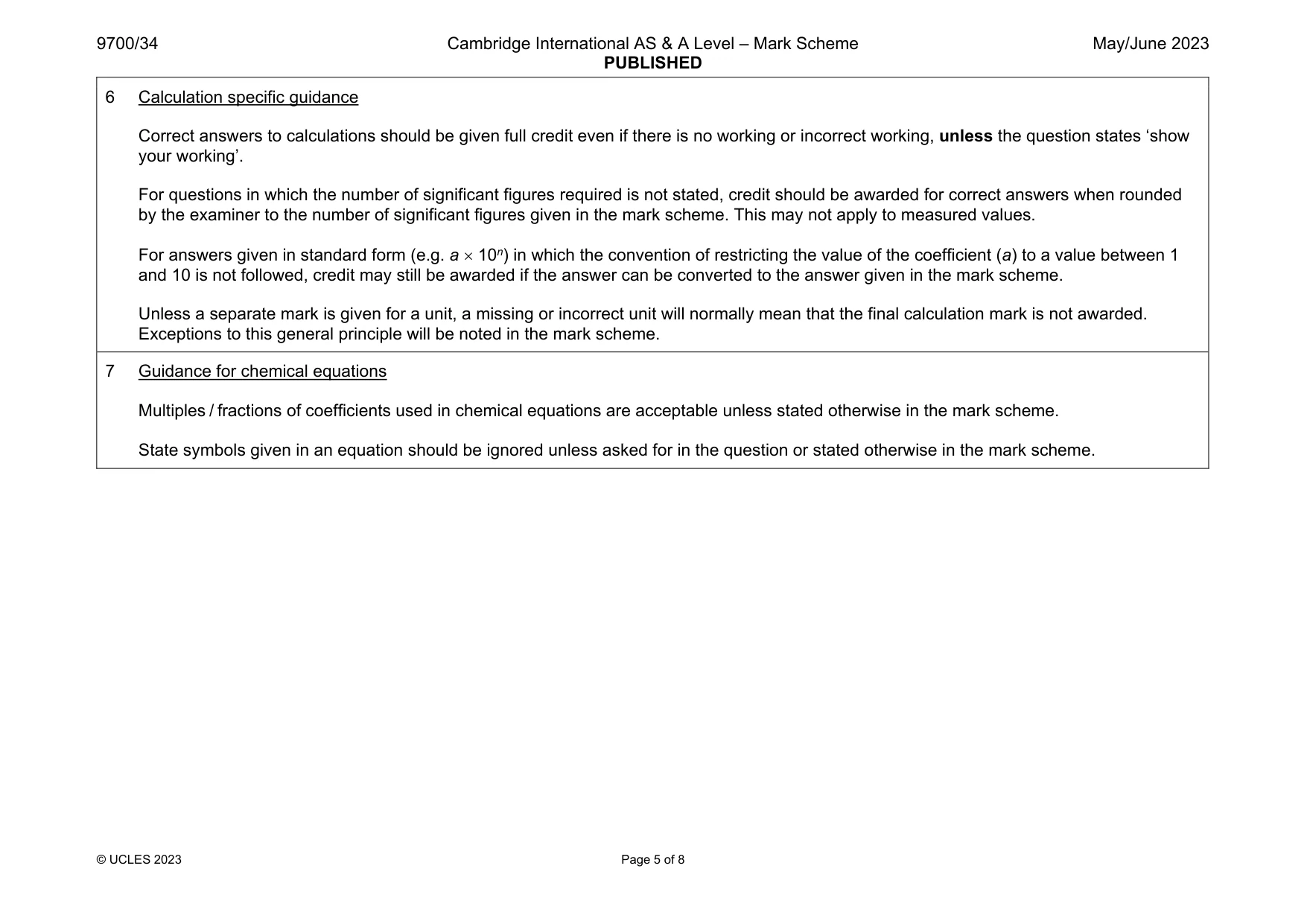
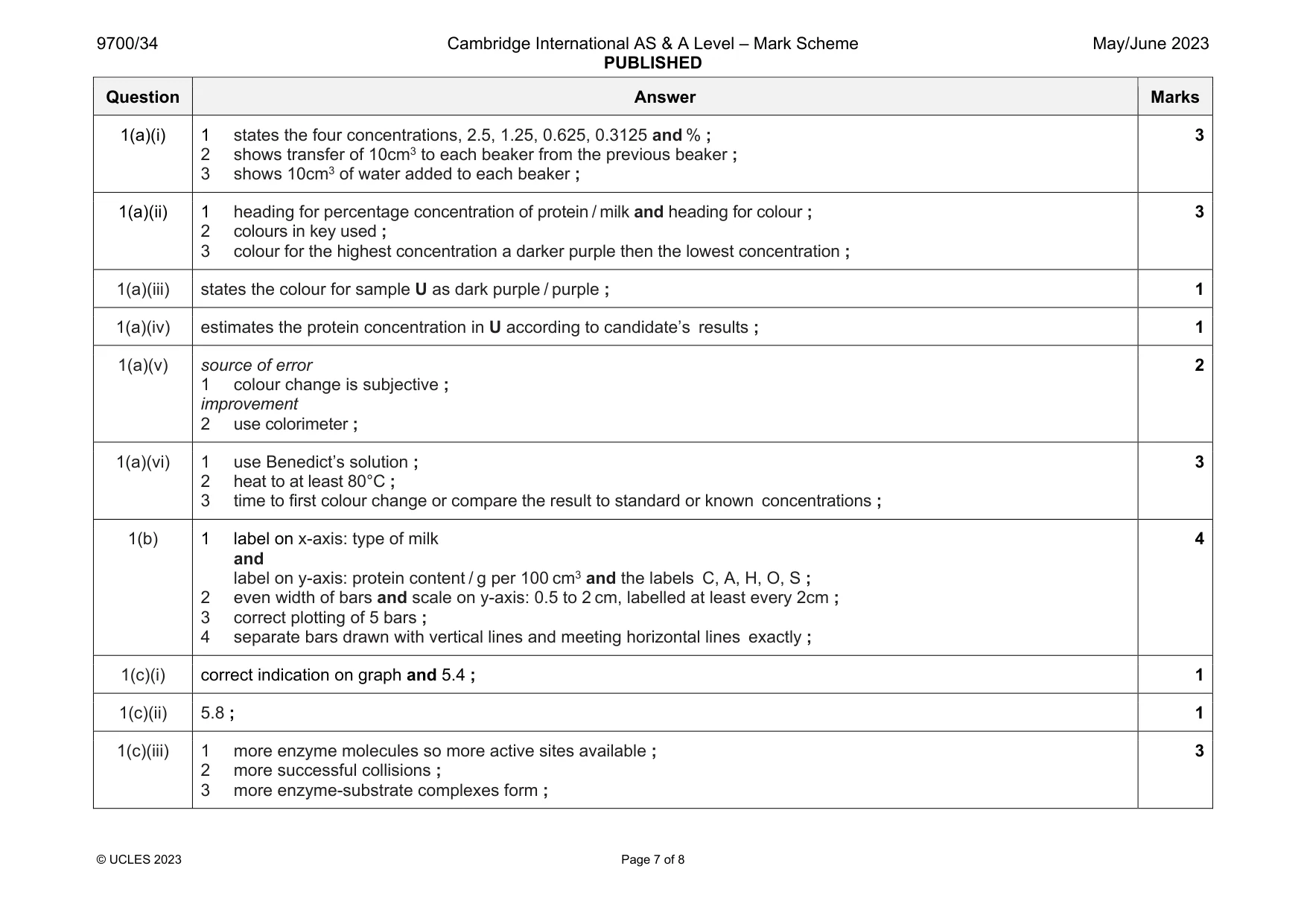
Mark scheme: 1(a)(i) 1 states the four concentrations, 2.5, 1.25, 0.625, 0.3125 and % ; 2 shows transfer of 10cm3 to each beaker from the previous beaker ; 3 shows 10cm3 of water added to each beaker ; 3 1(a)(ii) 1 heading for percentage concentration of protein / milk and heading for colour ; 2 colours in key used ; 3 colour for the highest concentration a darker purple then the lowest concentration ; 3 1(a)(iii) states the colour for sample U as dark purple / purple ; 1 1(a)(iv) estimates the protein concentration in U according to candidate’s results ; 1 1(a)(v) source of error 1 colour change is subjective ; improvement 2 use colorimeter ; 2 1(a)(vi) 1 use Benedict’s solution ; 2 heat to at least 80°C ; 3 time to first colour change or compare the result to standard or known concentrations ; 3 1(b) 1 label on x-axis: type of milk and label on y-axis: protein content / g per 100 cm3 and the labels C, A, H, O, S ; 2 even width of bars and scale on y-axis: 0.5 to 2 cm, labelled at least every 2cm ; 3 correct plotting of 5 bars ; 4 separate bars drawn with vertical lines and meeting horizontal lines exactly ; 4 1(c)(i) correct indication on graph and 5.4 ; 1 1(c)(ii) 5.8 ; 1 1(c)(iii) 1 more enzyme molecules so more active sites available ; 2 more successful collisions ; 3 more enzyme-substrate complexes form ; 3
Q2 · N1 is a slide of a stained transverse section through a plant stem
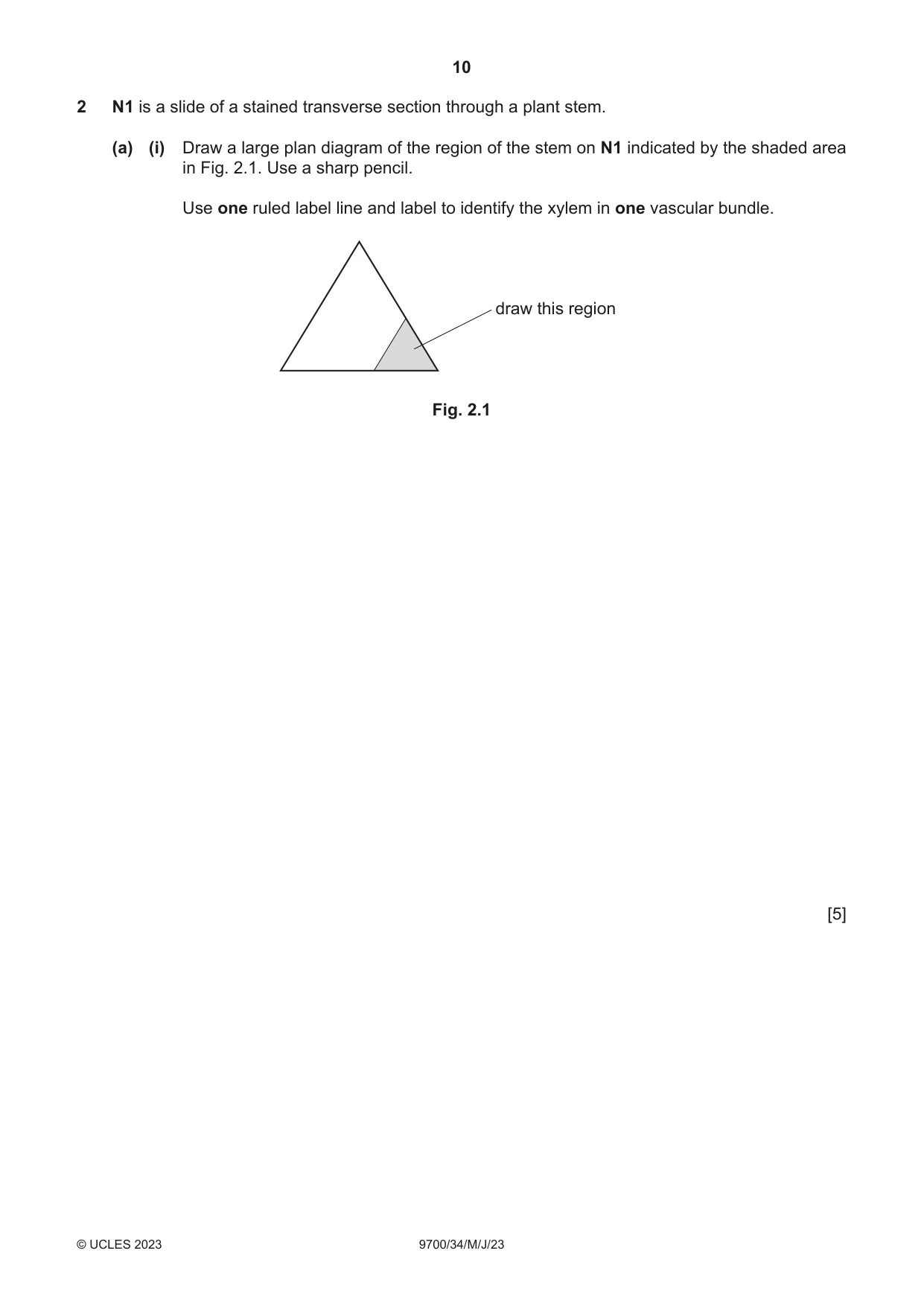
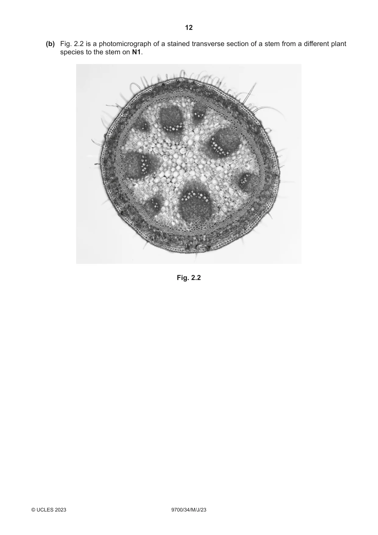
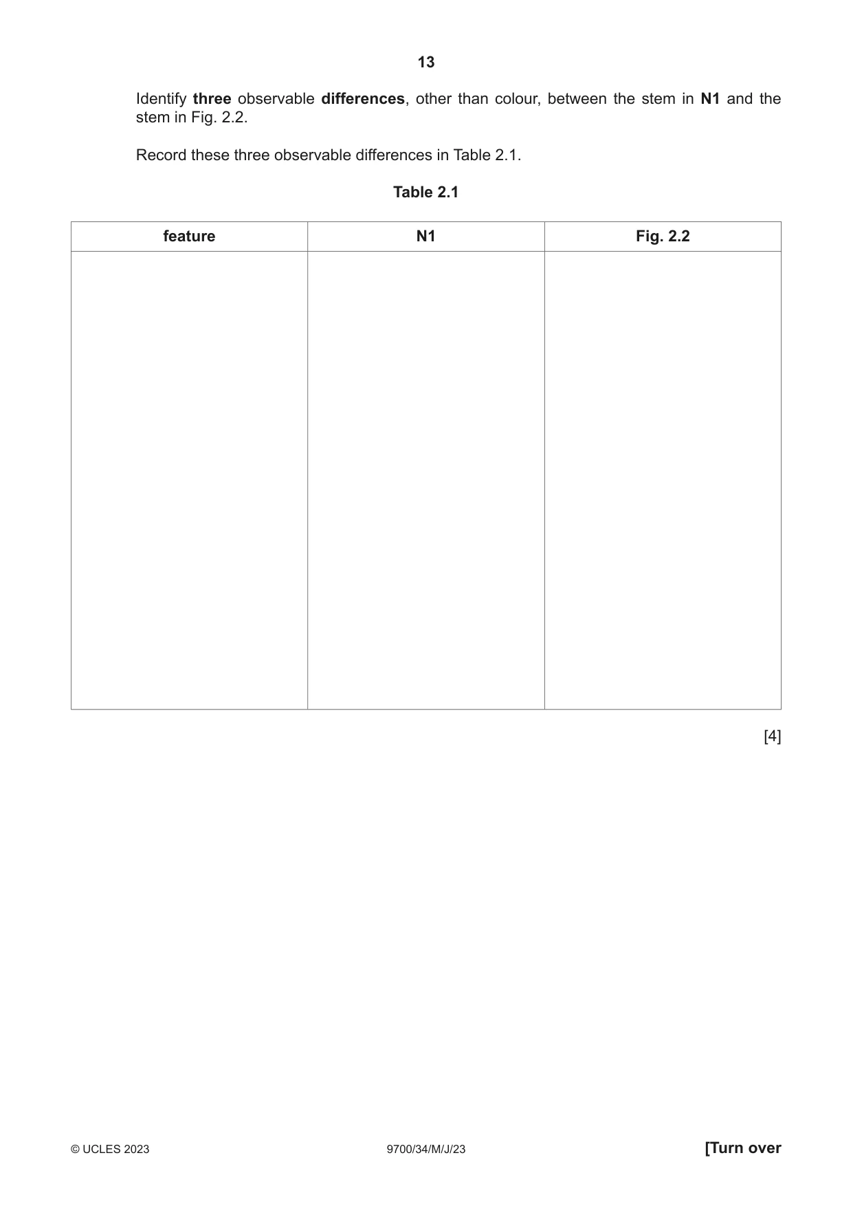
2 N1 is a slide of a stained transverse section through a plant stem. (a) (i) Draw a large plan diagram of the region of the stem on N1 indicated by the shaded area in Fig. 2.1. Use a sharp pencil. Use one ruled label line and label to identify the xylem in one vascular bundle. draw this region Fig. 2.1 [5] (ii) Observe the cells in the central tissue of the stem on N1. Select a group of four adjacent cells that make up this tissue. Each cell must touch at least two of the other cells. • Make a large drawing of this group of four cells. • Use one ruled label line and label to identify the cell wall of one cell. [5] (b) Fig. 2.2 is a photomicrograph of a stained transverse section of a stem from a different plant species to the stem on N1. Fig. 2.2
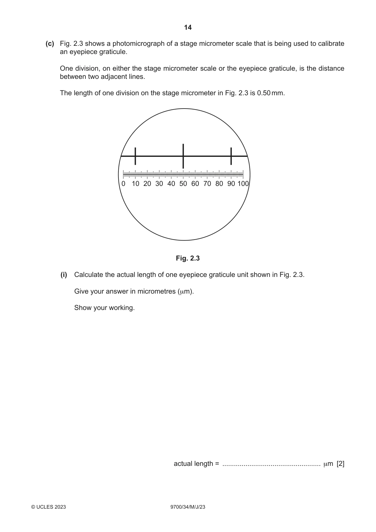
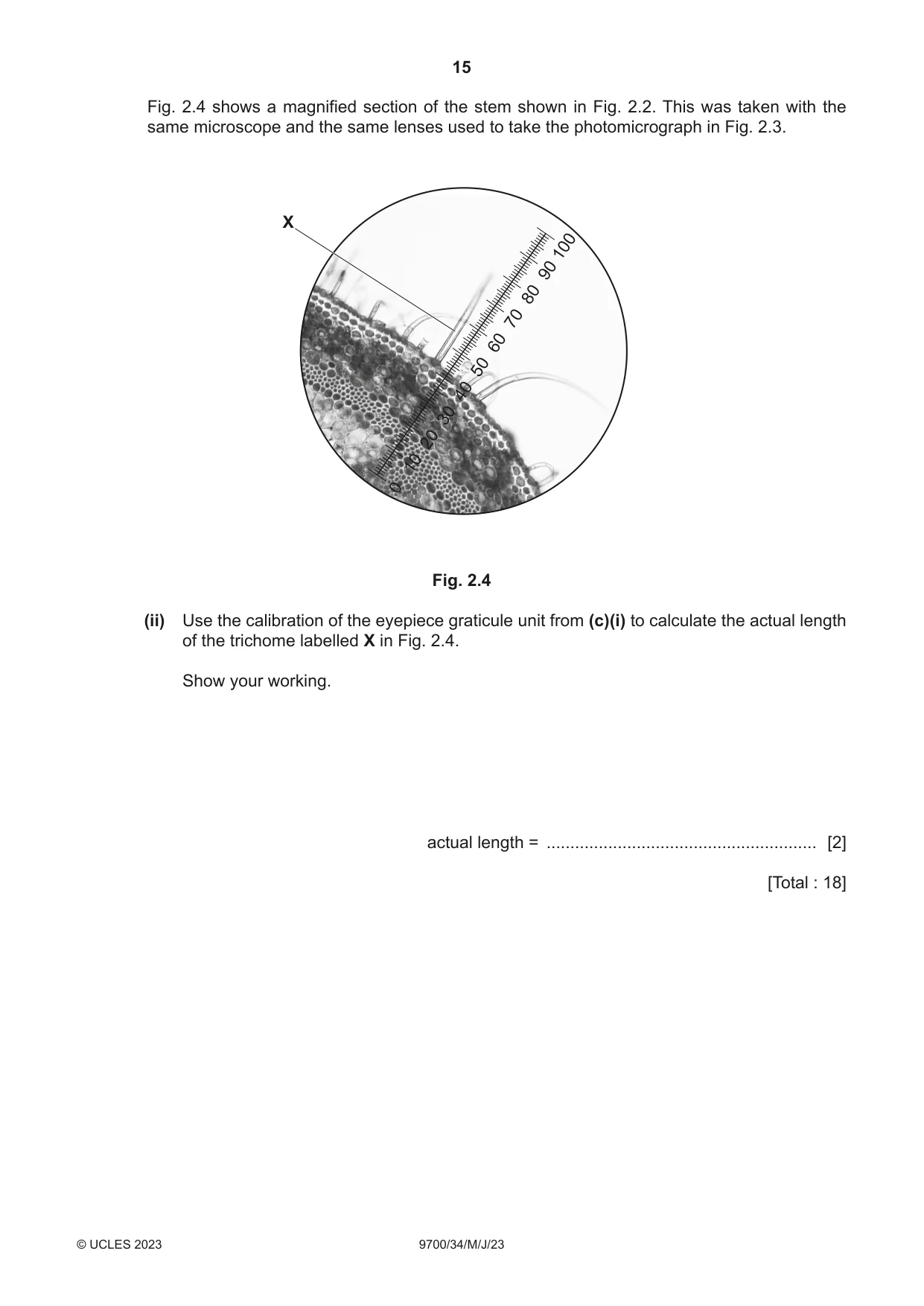
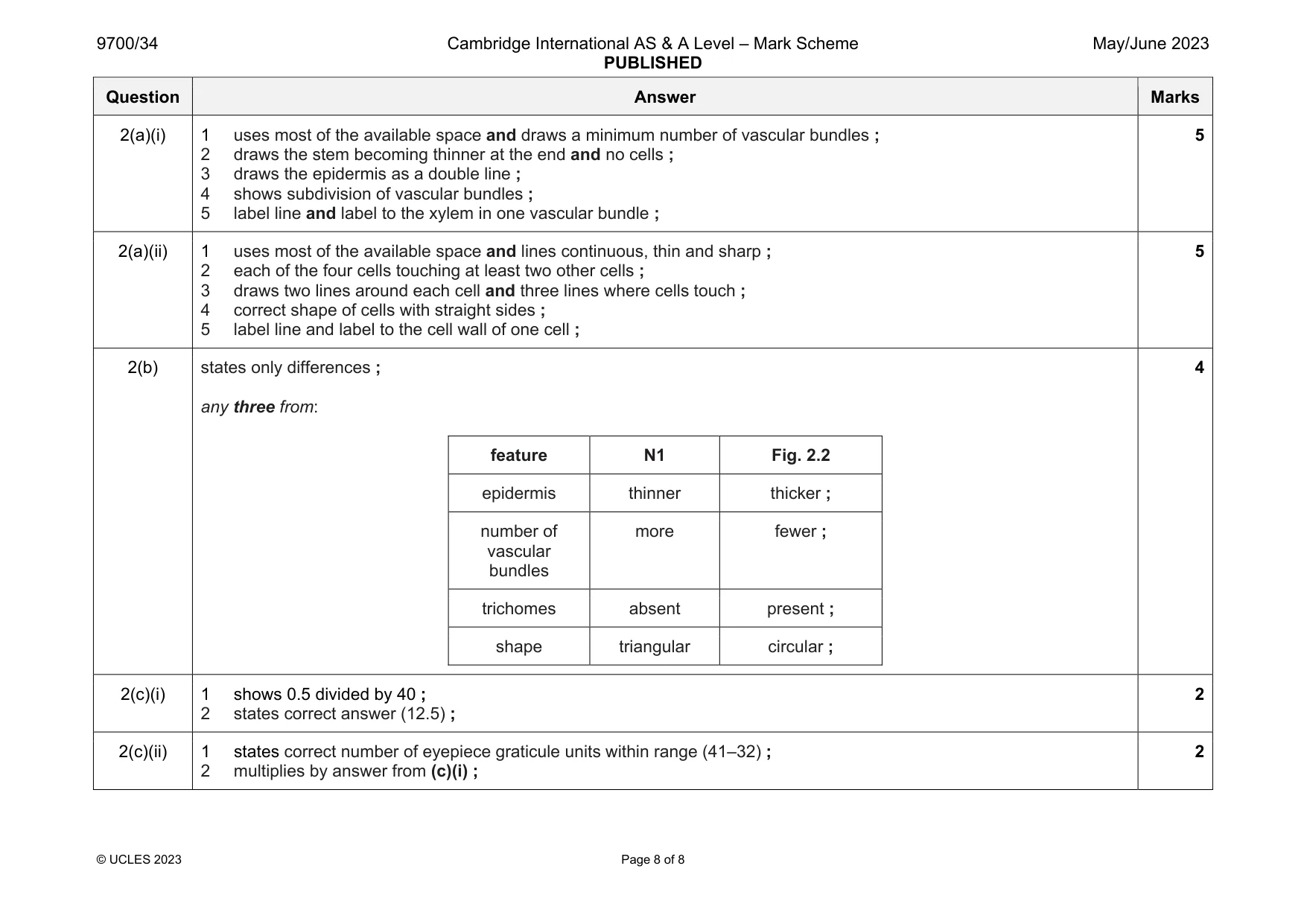
Mark scheme: 2(a)(i) 1 uses most of the available space and draws a minimum number of vascular bundles ; 2 draws the stem becoming thinner at the end and no cells ; 3 draws the epidermis as a double line ; 4 shows subdivision of vascular bundles ; 5 label line and label to the xylem in one vascular bundle ; 5 2(a)(ii) 1 uses most of the available space and lines continuous, thin and sharp ; 2 each of the four cells touching at least two other cells ; 3 draws two lines around each cell and three lines where cells touch ; 4 correct shape of cells with straight sides ; 5 label line and label to the cell wall of one cell ; 5 2(b) states only differences ; any three from: feature N1 Fig. 2.2 epidermis thinner thicker ; number of vascular bundles more fewer ; trichomes absent present ; shape triangular circular ; 4 2(c)(i) 1 shows 0.5 divided by 40 ; 2 states correct answer (12.5) ; 2 2(c)(ii) 1 states correct number of eyepiece graticule units within range (41–32) ; 2 multiplies by answer from (c)(i) ; 2
What was in this paper
The subtopics covered by these 2 questions, and how many questions each got. Open one in a new tab to see every Cambridge question on it.
What you needed in this session
Cambridge’s own grade thresholds for 2023 May/June, Paper 3 · Variant 4. A higher threshold means an easier paper — the bar moves with how the cohort did.