Cambridge A Level Biology 9700 — 2014 May/June Paper 3 · Variant 5
9700/35/M/J/14 · 40 marks · ≈45 min
The question paper and its mark scheme, free to read here and free to download. This is Cambridge’s own paper, exactly as it was sat.
Question paper12 pages

Mark scheme4 pages
Answers below. Sit the paper first if you are practising.
Paper as text
Question paper, page 1
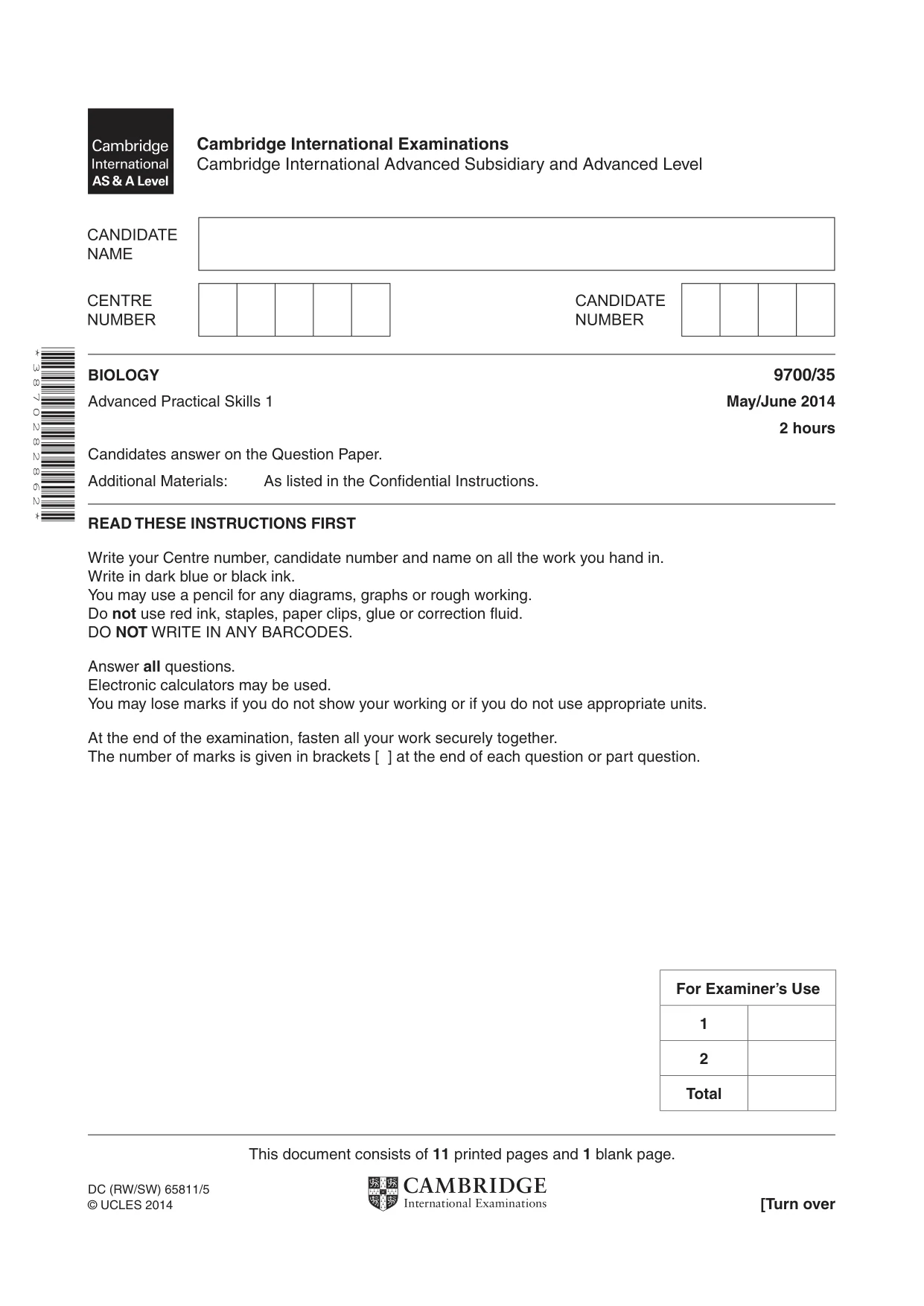
This document consists of 11 printed pages and 1 blank page. DC (RW/SW) 65811/5 © UCLES 2014 [Turn over Cambridge International Examinations Cambridge International Advanced Subsidiary and Advanced Level * 3 8 7 0 2 8 2 8 6 2 * BIOLOGY 9700/35 Advanced Practical Skills 1 May/June 2014 2 hours Candidates answer on the Question Paper. Additional Materials: As listed in the Confidential Instructions. READ THESE INSTRUCTIONS FIRST Write your Centre number, candidate number and name on all the work you hand in. Write in dark blue or black ink. You may use a pencil for any diagrams, graphs or rough working. Do not use red ink, staples, paper clips, glue or correction fluid. DO NOT WRITE IN ANY BARCODES. Answer all questions. Electronic calculators may be used. You may lose marks if you do not show your working or if you do not use appropriate units. At the end of the examination, fasten all your work securely together. The number of marks is given in brackets [ ] at the end of each question or part question. For Examiner’s Use 1 2 Total
Question paper, page 2
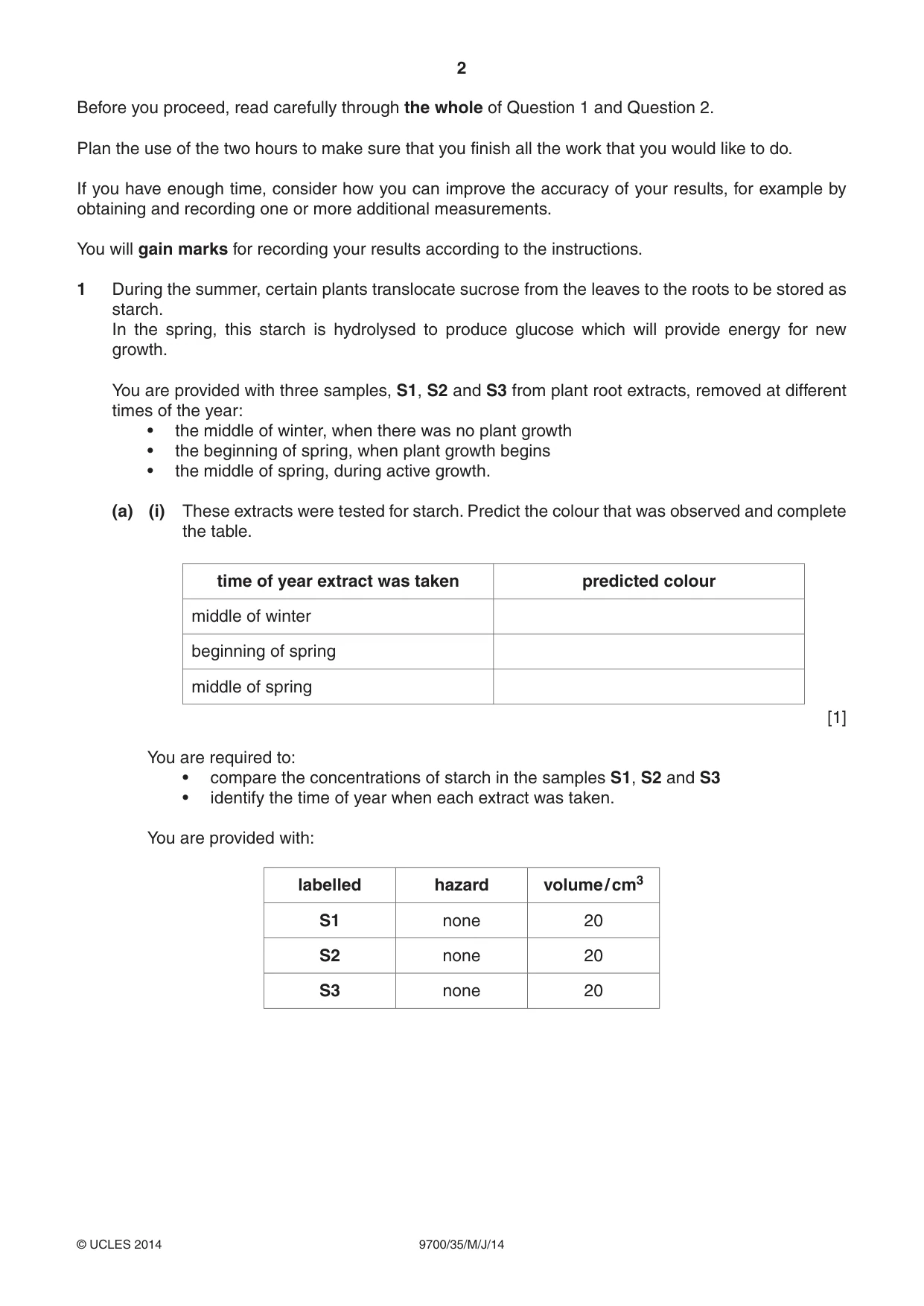
2 9700/35/M/J/14 © UCLES 2014 Before you proceed, read carefully through the whole of Question 1 and Question 2. Plan the use of the two hours to make sure that you finish all the work that you would like to do. If you have enough time, consider how you can improve the accuracy of your results, for example by obtaining and recording one or more additional measurements. You will gain marks for recording your results according to the instructions. 1 During the summer, certain plants translocate sucrose from the leaves to the roots to be stored as starch. In the spring, this starch is hydrolysed to produce glucose which will provide energy for new growth. You are provided with three samples, S1, S2 and S3 from plant root extracts, removed at different times of the year: • the middle of winter, when there was no plant growth • the beginning of spring, when plant growth begins • the middle of spring, during active growth. (a) (i) These extracts were tested for starch. Predict the colour that was observed and complete the table. time of year extract was taken predicted colour middle of winter beginning of spring middle of spring [1] You are required to: • compare the concentrations of starch in the samples S1, S2 and S3 • identify the time of year when each extract was taken. You are provided with: labelled hazard volume / cm3 S1 none 20 S2 none 20 S3 none 20
Question paper, page 3
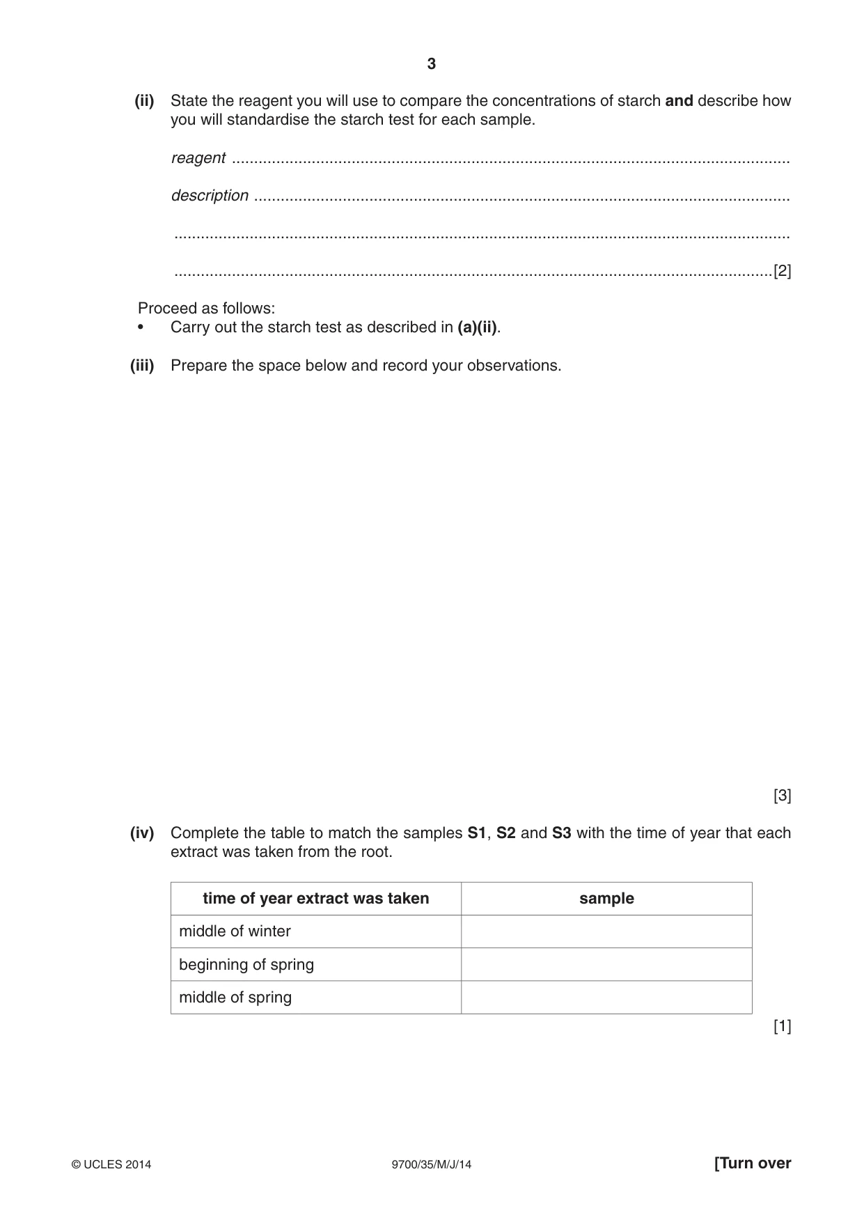
3 9700/35/M/J/14 © UCLES 2014 [Turn over (ii) State the reagent you will use to compare the concentrations of starch and describe how you will standardise the starch test for each sample. reagent … description … … …[2] Proceed as follows: • Carry out the starch test as described in (a)(ii). (iii) Prepare the space below and record your observations. [3] (iv) Complete the table to match the samples S1, S2 and S3 with the time of year that each extract was taken from the root. time of year extract was taken sample middle of winter beginning of spring middle of spring [1]
Question paper, page 4
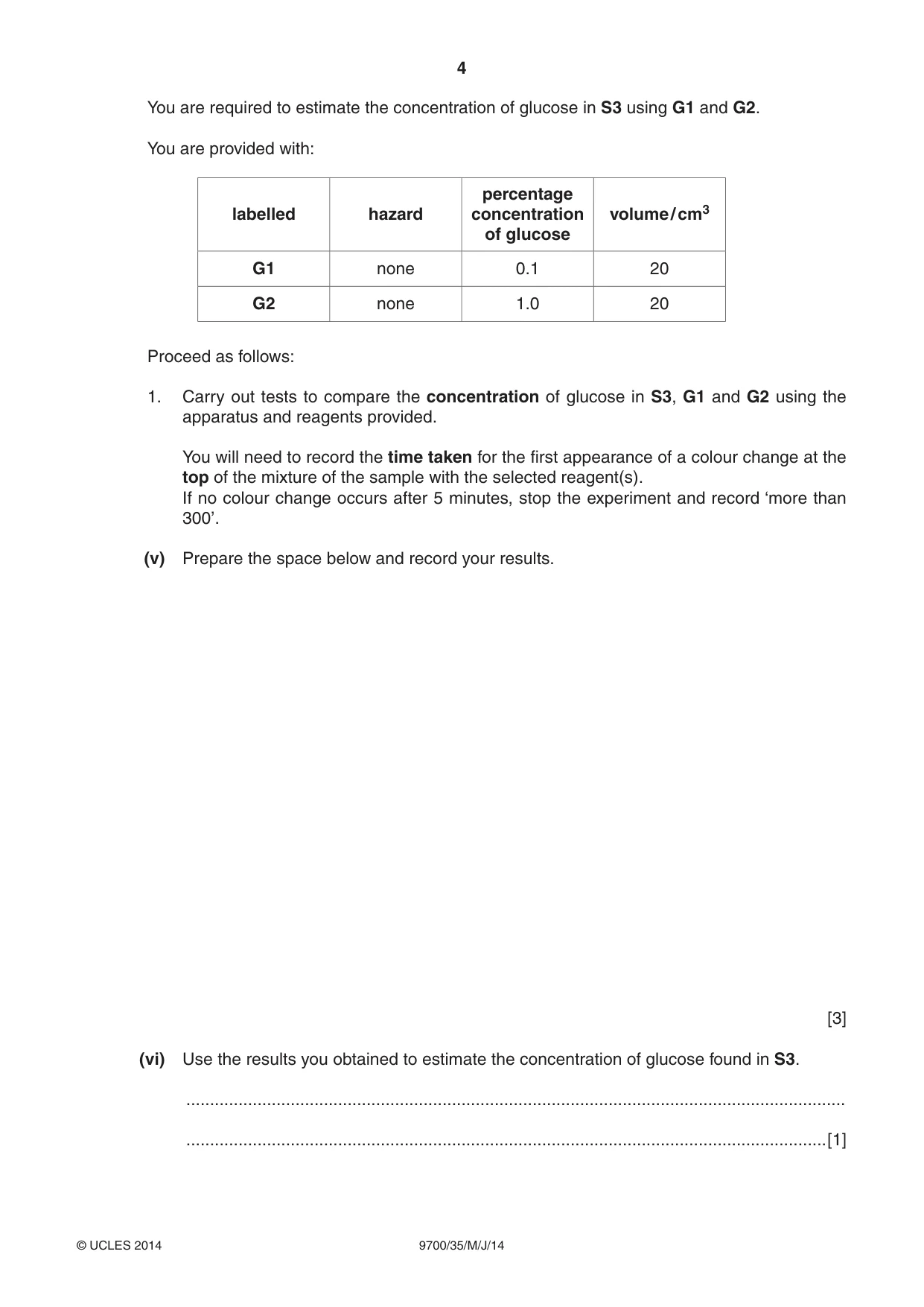
4 9700/35/M/J/14 © UCLES 2014 You are required to estimate the concentration of glucose in S3 using G1 and G2. You are provided with: labelled hazard percentage concentration of glucose volume / cm3 G1 none 0.1 20 G2 none 1.0 20 Proceed as follows: 1. Carry out tests to compare the concentration of glucose in S3, G1 and G2 using the apparatus and reagents provided. You will need to record the time taken for the first appearance of a colour change at the top of the mixture of the sample with the selected reagent(s). If no colour change occurs after 5 minutes, stop the experiment and record ‘more than 300’. (v) Prepare the space below and record your results. [3] (vi) Use the results you obtained to estimate the concentration of glucose found in S3. … …[1]
Question paper, page 5
5 9700/35/M/J/14 © UCLES 2014 [Turn over (vii) Suggest how you would modify this investigation to obtain a more accurate estimate of the glucose concentration in S3. … … … … … … …[3] (viii) A systematic error occurs when apparatus with scales are used, since the scales may be slightly different. For example, when measuring the same line, two rulers may give different lengths. However, as long as the same ruler is used for all the measurements, the trend is not affected because the error is consistent. State one piece of apparatus used in this investigation that may have a systematic error. Suggest whether this affected your results and give a reason for your answer. apparatus … effect and reason … …[1]
Question paper, page 6
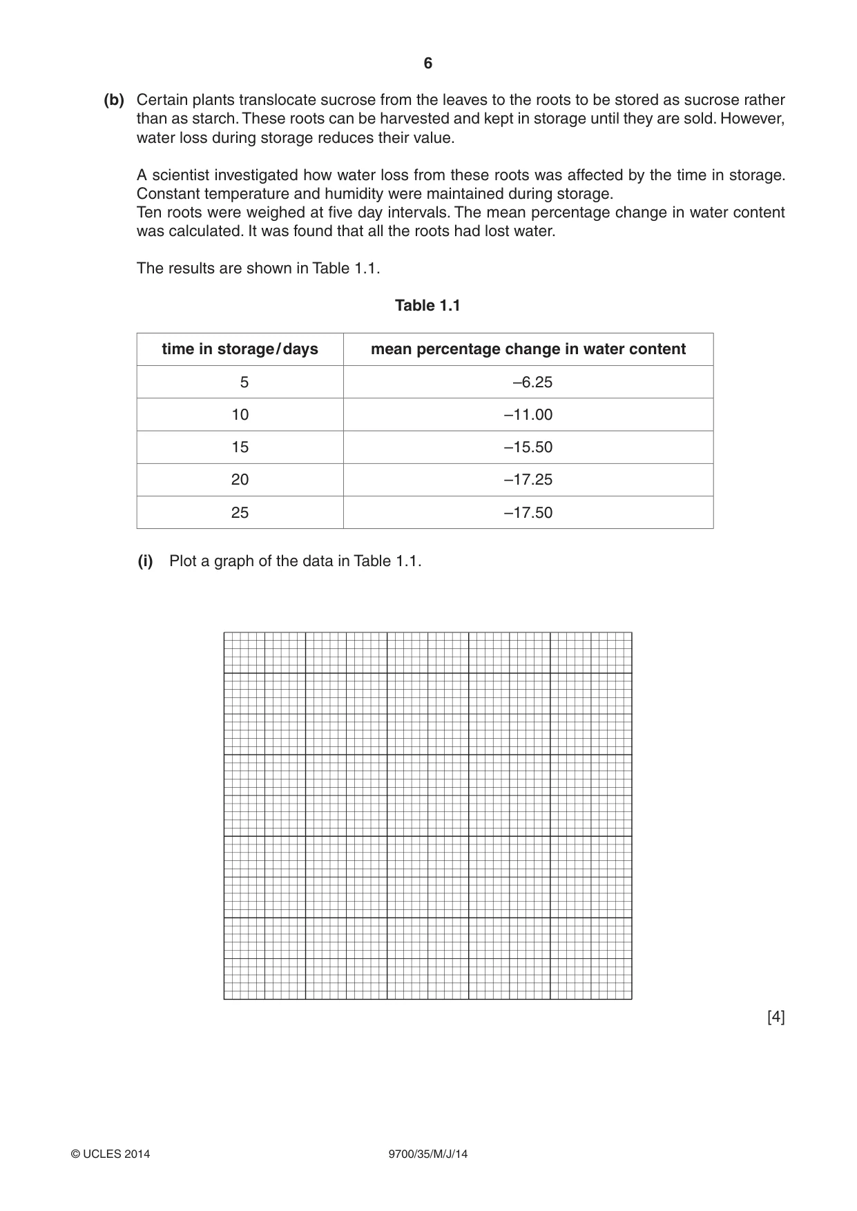
6 9700/35/M/J/14 © UCLES 2014 (b) Certain plants translocate sucrose from the leaves to the roots to be stored as sucrose rather than as starch. These roots can be harvested and kept in storage until they are sold. However, water loss during storage reduces their value. A scientist investigated how water loss from these roots was affected by the time in storage. Constant temperature and humidity were maintained during storage. Ten roots were weighed at five day intervals. The mean percentage change in water content was calculated. It was found that all the roots had lost water. The results are shown in Table 1.1. Table 1.1 time in storage / days mean percentage change in water content 5 –6.25 10 –11.00 15 –15.50 20 –17.25 25 –17.50 (i) Plot a graph of the data in Table 1.1. [4]
Question paper, page 7
7 9700/35/M/J/14 © UCLES 2014 [Turn over (ii) Ten roots were stored for 5 days, and another ten roots were stored for 20 days. Pieces of root from the sample at 5 days and from the sample at 20 days were removed, weighed and soaked in dilute sucrose solution for 24 hours. The pieces were then reweighed. State which sample would have the highest percentage gain in mass after soaking in the dilute sucrose solution. Explain your answer using your knowledge of water potential. sample … explanation … … … …[2] [Total: 21]
Question paper, page 8
8 9700/35/M/J/14 © UCLES 2014 2 The eyepiece graticule scale in your microscope may be used to measure the actual length of the layers of tissue or cells, if the scale has been calibrated against a stage micrometer. However, to help draw the correct shape and proportion of tissues, as in (a), it is not necessary to calibrate the eyepiece graticule scale. K1 and K2 are slides showing stained transverse sections through blood vessels. (a) Draw large plan diagrams of two different types of blood vessel shown in K1 and K2. On your diagram, use ruled label lines and labels to describe (annotate) one observable difference in the layer lining the inside of each vessel. [5]
Question paper, page 9
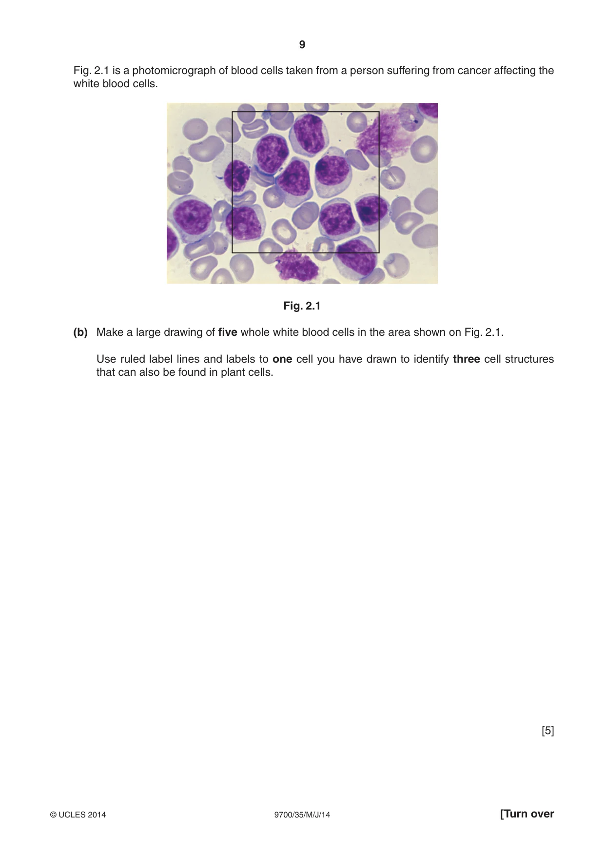
9 9700/35/M/J/14 © UCLES 2014 [Turn over Fig. 2.1 is a photomicrograph of blood cells taken from a person suffering from cancer affecting the white blood cells. Fig. 2.1 (b) Make a large drawing of five whole white blood cells in the area shown on Fig. 2.1. Use ruled label lines and labels to one cell you have drawn to identify three cell structures that can also be found in plant cells. [5]
Question paper, page 10
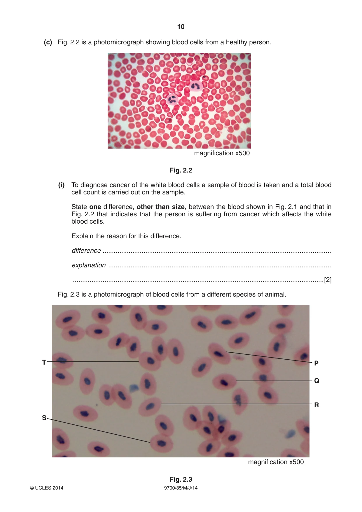
10 9700/35/M/J/14 © UCLES 2014 (c) Fig. 2.2 is a photomicrograph showing blood cells from a healthy person. magnification x500 Fig. 2.2 (i) To diagnose cancer of the white blood cells a sample of blood is taken and a total blood cell count is carried out on the sample. State one difference, other than size, between the blood shown in Fig. 2.1 and that in Fig. 2.2 that indicates that the person is suffering from cancer which affects the white blood cells. Explain the reason for this difference. difference … explanation … …[2] Fig. 2.3 is a photomicrograph of blood cells from a different species of animal. magnification x500 T P Q R S Fig. 2.3
Question paper, page 11
11 9700/35/M/J/14 © UCLES 2014 (ii) A student measured the longest length of five red blood cells P, Q, R, S and T shown in Fig. 2.3 and found the mean actual length of the red blood cells. Use the magnification to calculate the mean actual length, in μm, of the red blood cells using the five labelled red blood cells in Fig. 2.3. You may lose marks if you do not show your working or if you do not use appropriate units. mean actual length …μm [4] (d) Prepare the space below so that it is suitable for you to record the observable differences between the red blood cells in Fig. 2.2 and Fig. 2.3. Record your observations in the space you have prepared. [3] [Total: 19]
Question paper, page 12
12 9700/35/M/J/14 © UCLES 2014 Copyright Acknowledgements: Fig. 2.1 © BONHOMME, ISM/SCIENCE PHOTO LIBRARY. Fig. 2.2 © ERIC GRAVE/SCIENCE PHOTO LIBRARY. Permission to reproduce items where third-party owned material protected by copyright is included has been sought and cleared where possible. Every reasonable effort has been made by the publisher (UCLES) to trace copyright holders, but if any items requiring clearance have unwittingly been included, the publisher will be pleased to make amends at the earliest possible opportunity. Cambridge International Examinations is part of the Cambridge Assessment Group. Cambridge Assessment is the brand name of University of Cambridge Local Examinations Syndicate (UCLES), which is itself a department of the University of Cambridge. BLANK PAGE
Mark scheme, page 1
CAMBRIDGE INTERNATIONAL EXAMINATIONS GCE Advanced Subsidiary Level and GCE Advanced Level MARK SCHEME for the May/June 2014 series 9700 BIOLOGY 9700/35 Paper 35 (Advanced Practical Skills 1), maximum raw mark 40 This mark scheme is published as an aid to teachers and candidates, to indicate the requirements of the examination. It shows the basis on which Examiners were instructed to award marks. It does not indicate the details of the discussions that took place at an Examiners’ meeting before marking began, which would have considered the acceptability of alternative answers. Mark schemes should be read in conjunction with the question paper and the Principal Examiner Report for Teachers. Cambridge will not enter into discussions about these mark schemes. Cambridge is publishing the mark schemes for the May/June 2014 series for most IGCSE, GCE Advanced Level and Advanced Subsidiary Level components and some Ordinary Level components.
Mark scheme, page 2
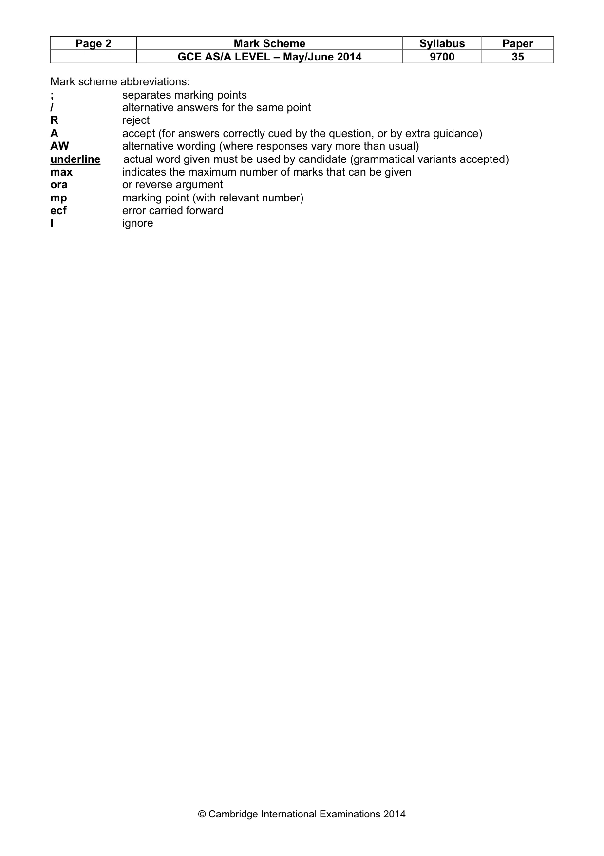
Page 2 Mark Scheme Syllabus Paper GCE AS/A LEVEL – May/June 2014 9700 35 © Cambridge International Examinations 2014 Mark scheme abbreviations: ; separates marking points / alternative answers for the same point R reject A accept (for answers correctly cued by the question, or by extra guidance) AW alternative wording (where responses vary more than usual) underline actual word given must be used by candidate (grammatical variants accepted) max indicates the maximum number of marks that can be given ora or reverse argument mp marking point (with relevant number) ecf error carried forward I ignore
Mark scheme, page 3
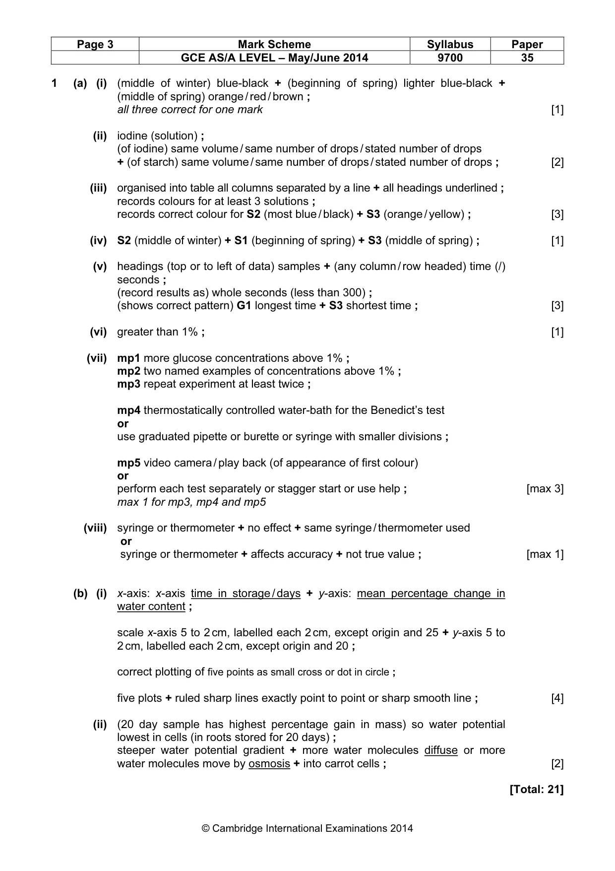
Page 3 Mark Scheme Syllabus Paper GCE AS/A LEVEL – May/June 2014 9700 35 © Cambridge International Examinations 2014 1 (a) (i) (middle of winter) blue-black + (beginning of spring) lighter blue-black + (middle of spring) orange / red / brown ; all three correct for one mark [1] (ii) iodine (solution) ; (of iodine) same volume / same number of drops / stated number of drops + (of starch) same volume / same number of drops / stated number of drops ; [2] (iii) organised into table all columns separated by a line + all headings underlined ; records colours for at least 3 solutions ; records correct colour for S2 (most blue / black) + S3 (orange / yellow) ; [3] (iv) S2 (middle of winter) + S1 (beginning of spring) + S3 (middle of spring) ; [1] (v) headings (top or to left of data) samples + (any column / row headed) time (/) seconds ; (record results as) whole seconds (less than 300) ; (shows correct pattern) G1 longest time + S3 shortest time ; [3] (vi) greater than 1% ; [1] (vii) mp1 more glucose concentrations above 1% ; mp2 two named examples of concentrations above 1% ; mp3 repeat experiment at least twice ; mp4 thermostatically controlled water-bath for the Benedict’s test or use graduated pipette or burette or syringe with smaller divisions ; mp5 video camera / play back (of appearance of first colour) or perform each test separately or stagger start or use help ; [max 3] max 1 for mp3, mp4 and mp5 (viii) syringe or thermometer + no effect + same syringe / thermometer used or syringe or thermometer + affects accuracy + not true value ; [max 1] (b) (i) x-axis: x-axis time in storage / days + y-axis: mean percentage change in water content ; scale x-axis 5 to 2 cm, labelled each 2 cm, except origin and 25 + y-axis 5 to 2 cm, labelled each 2 cm, except origin and 20 ; correct plotting of five points as small cross or dot in circle ; five plots + ruled sharp lines exactly point to point or sharp smooth line ; [4] (ii) (20 day sample has highest percentage gain in mass) so water potential lowest in cells (in roots stored for 20 days) ; steeper water potential gradient + more water molecules diffuse or more water molecules move by osmosis + into carrot cells ; [2] [Total: 21]
Mark scheme, page 4
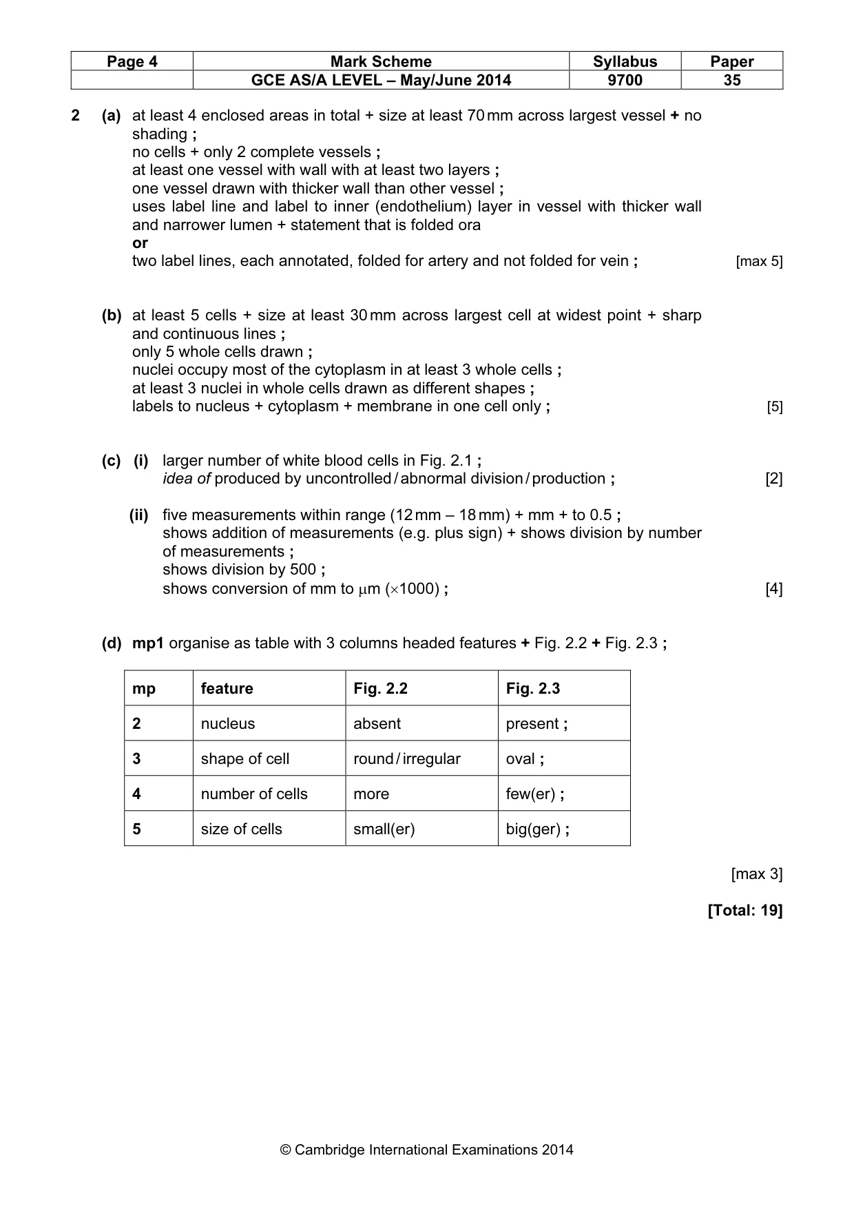
Page 4 Mark Scheme Syllabus Paper GCE AS/A LEVEL – May/June 2014 9700 35 © Cambridge International Examinations 2014 2 (a) at least 4 enclosed areas in total + size at least 70 mm across largest vessel + no shading ; no cells + only 2 complete vessels ; at least one vessel with wall with at least two layers ; one vessel drawn with thicker wall than other vessel ; uses label line and label to inner (endothelium) layer in vessel with thicker wall and narrower lumen + statement that is folded ora or two label lines, each annotated, folded for artery and not folded for vein ; [max 5] (b) at least 5 cells + size at least 30 mm across largest cell at widest point + sharp and continuous lines ; only 5 whole cells drawn ; nuclei occupy most of the cytoplasm in at least 3 whole cells ; at least 3 nuclei in whole cells drawn as different shapes ; labels to nucleus + cytoplasm + membrane in one cell only ; [5] (c) (i) larger number of white blood cells in Fig. 2.1 ; idea of produced by uncontrolled / abnormal division / production ; [2] (ii) five measurements within range (12 mm – 18 mm) + mm + to 0.5 ; shows addition of measurements (e.g. plus sign) + shows division by number of measurements ; shows division by 500 ; shows conversion of mm to µm (×1000) ; [4] (d) mp1 organise as table with 3 columns headed features + Fig. 2.2 + Fig. 2.3 ; mp feature Fig. 2.2 Fig. 2.3 2 nucleus absent present ; 3 shape of cell round / irregular oval ; 4 number of cells more few(er) ; 5 size of cells small(er) big(ger) ; [max 3] [Total: 19]
What you needed in this session
Cambridge’s own grade thresholds for 2014 May/June, Paper 3 · Variant 5. A higher threshold means an easier paper — the bar moves with how the cohort did.